Possible Combined Central and Peripheral Demyelination Presenting as Optic Neuritis, Cervical Myelitis, and Demyelinating Polyneuropathy with Marked Nerve Hypertrophy
- PMID: 29540658
- PMCID: PMC5891529
- DOI: 10.2169/internalmedicine.7153-16
Possible Combined Central and Peripheral Demyelination Presenting as Optic Neuritis, Cervical Myelitis, and Demyelinating Polyneuropathy with Marked Nerve Hypertrophy
Abstract
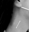
A 27-year-old woman with optic neuritis and cervical myelitis developed hypertrophic demyelinating polyneuropathy. It was hypothesized that the diagnosis was combined central and peripheral demyelination. A hypertrophic nerve was observed subcutaneously, and magnetic resonance imaging demonstrated marked hypertrophy of the nerve roots. The patient was negative for anti-aquaporin 4 antibodies. Her anti-neurofascin 155 antibody levels was slightly elevated, but it was not definitely positive. Pulsed steroid therapy and the administration of immunoglobulin ameliorated her symptoms. Molecules in both the peripheral and central nervous systems might be target antigens, but further investigations will be needed to clarify the precise pathogenic mechanisms.
Keywords: chronic inflammatory demyelinating polyneuropathy; combined central and peripheral demyelination; multiple sclerosis; neurofascin.
Figures


Similar articles
-
Shifting borders, crossing boundaries: The case of combined central and peripheral demyelination.Mult Scler. 2018 Apr;24(4):550-551. doi: 10.1177/1352458517726386. Epub 2017 Aug 10. Mult Scler. 2018. PMID: 28795614
-
Optic neuritis and multiple sclerosis.Curr Opin Neurol. 2008 Feb;21(1):16-21. doi: 10.1097/WCO.0b013e3282f419ca. Curr Opin Neurol. 2008. PMID: 18180647 Review.
-
Acute simultaneous development of brain tumour-like lesion and demyelinating polyneuropathy in a patient with chronic relapsing myelitis.Mult Scler. 2018 Apr;24(4):546-550. doi: 10.1177/1352458517714610. Epub 2017 Aug 10. Mult Scler. 2018. PMID: 28795610
-
Fulminant Central Plus Peripheral Nervous System Demyelination without Antibodies to Neurofascin.Can J Neurol Sci. 2016 Jan;43(1):149-56. doi: 10.1017/cjn.2015.238. Epub 2015 Aug 14. Can J Neurol Sci. 2016. PMID: 26271726
-
The limited demyelinating diseases: the voyage of optic neuritis and transverse myelitis to multiple sclerosis and neuromyelitis.Expert Rev Neurother. 2011 Mar;11(3):451-62. doi: 10.1586/ern.11.6. Expert Rev Neurother. 2011. PMID: 21375450 Review.
Cited by
-
Immune-mediated insights into clinical and specific autoantibodies in acute and chronic immune-mediated nodo-paranodopathies.Arq Neuropsiquiatr. 2025 Apr;83(4):1-6. doi: 10.1055/s-0045-1805073. Epub 2025 Mar 19. Arq Neuropsiquiatr. 2025. PMID: 40107278 Free PMC article. Review.
-
Is MS affecting the CNS only? Lessons from clinic to myelin pathophysiology.Neurol Neuroimmunol Neuroinflamm. 2020 Nov 24;8(1):e914. doi: 10.1212/NXI.0000000000000914. Print 2021 Jan. Neurol Neuroimmunol Neuroinflamm. 2020. PMID: 33234720 Free PMC article. Review.
-
Concurrent leukoencephalomyelitis and polyneuritis in a Maltese terrier: resembling combined central and peripheral demyelination in humans.J Vet Med Sci. 2019 Oct 10;81(9):1373-1378. doi: 10.1292/jvms.18-0696. Epub 2019 Jul 30. J Vet Med Sci. 2019. PMID: 31366813 Free PMC article.
-
MOG antibodies in combined central and peripheral demyelination syndromes.Neurol Neuroimmunol Neuroinflamm. 2018 Sep 11;5(6):e503. doi: 10.1212/NXI.0000000000000503. eCollection 2018 Nov. Neurol Neuroimmunol Neuroinflamm. 2018. PMID: 30246057 Free PMC article. No abstract available.
References
-
- Ro YI, Alexander CB, Oh SJ. Multiple sclerosis and hypertrophic demyelinating peripheral neuropathy. Muscle Nerve 6: 312-316, 1983. - PubMed
-
- Nonaka T, Fujimoto T, Eguchi K, Fukuda Y, Yoshimura T. Fujimoto T. Eguchi k. Fukuda Y. Yoshimura T. A case of combined and peripheral demyelination. Rinsho Shinkeigaku (Clin Neurol) 55: 389-394, 2015. (in Japanese, Abstract in English). - PubMed
-
- Butzkueven H, O'Brien TJ, Sedal L. Combined peripheral nerve and central nervous system demyelination in a patient with chronic inflammatory demyelinating polyneuropathy. J Clin Neurosci 6: 358-360, 1996. - PubMed
-
- Kawamura N, Yamasaki R, Yonekawa T, et al. . Anti-neurofascin antibody in patients with combined central and peripheral demyelination. Neurology 81: 714-722, 2013. - PubMed
-
- Yamasaki R. Anti-neurofascin antibody in combined central and peripheral demyelination. Clin Exp Neuroimmunol 4: 68-75, 2013.
Publication types
MeSH terms
Substances
LinkOut - more resources
Full Text Sources
Other Literature Sources
Medical

