. 2018 Mar 14;9(3):411.
doi: 10.1038/s41419-018-0450-z.

miR-381-3p knockdown improves intestinal epithelial proliferation and barrier function after intestinal ischemia/reperfusion injury by targeting nurr1

Affiliations

miR-381-3p knockdown improves intestinal epithelial proliferation and barrier function after intestinal ischemia/reperfusion injury by targeting nurr1

Liwei Liu et al. Cell Death Dis. .

Abstract

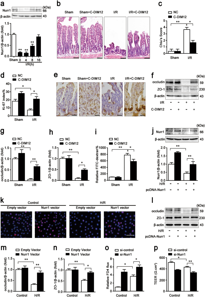
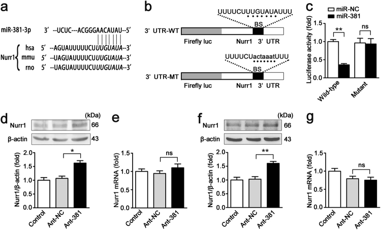
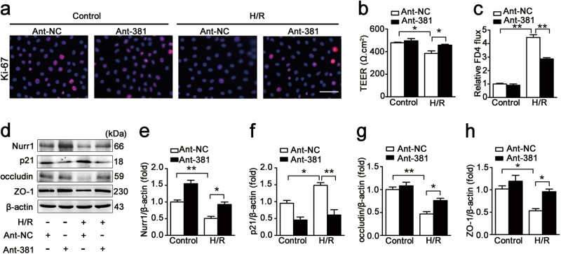
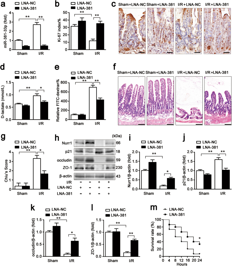
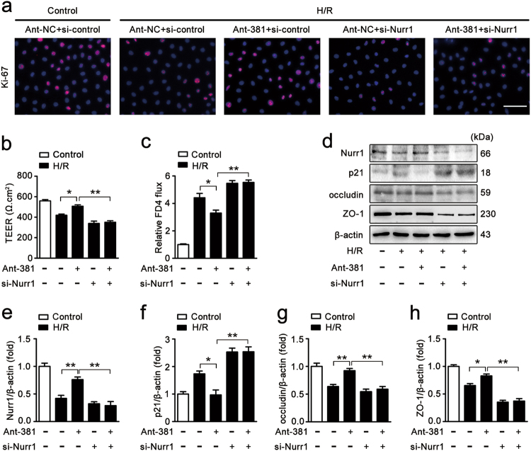
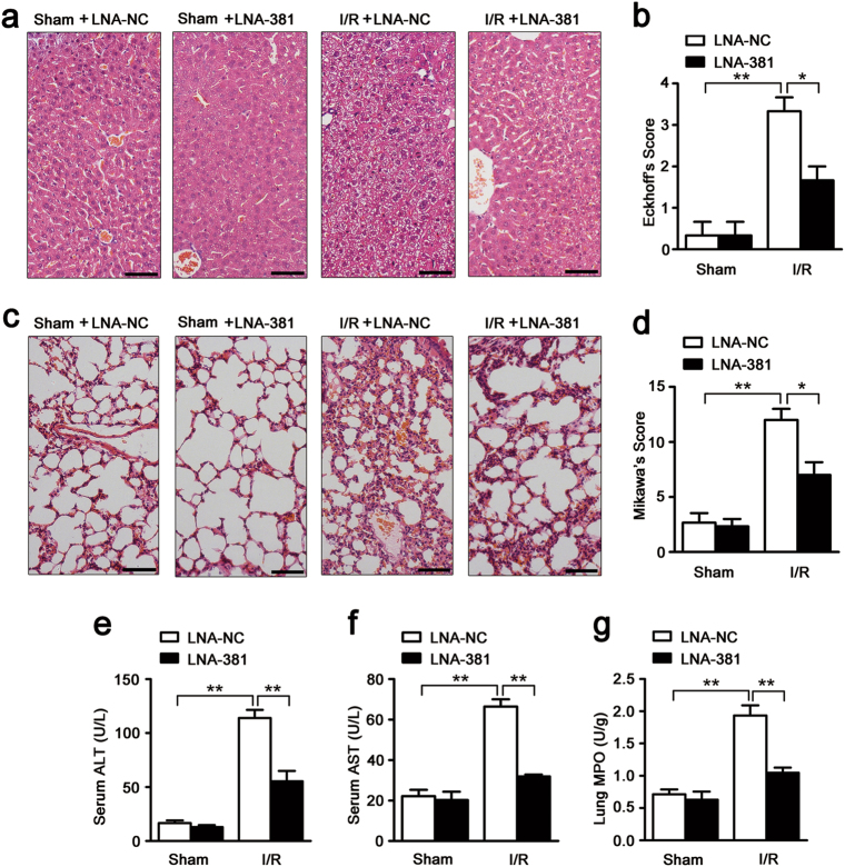
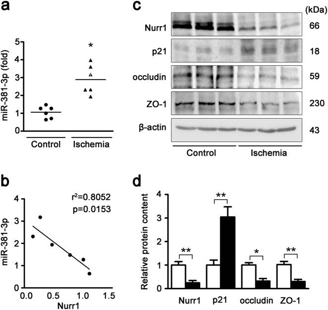
Impairment in gut barrier function induced by intestinal ischemia/reperfusion (I/R) injury is associated with high morbidity and mortality. Intestinal barrier function requires the tight coordination of epithelial migration, proliferation and differentiation. We previously observed that nuclear receptor-related protein 1 (nurr1)-mediated proliferative pathway was impaired in intestinal I/R injury. Here, we aimed to assess the effect of nurr1 on intestinal barrier function and to evaluate microRNA (miRNA)-nurr1-mediated restoration of intestinal barrier function in intestinal I/R injury. We induced an in vivo intestinal I/R injury mouse model by clamping and then releasing the superior mesenteric artery. We also performed an in vitro study in which we exposed Caco-2 and IEC-6 cells to hypoxia/reoxygenation (H/R) conditions to stimulate intestinal I/R injury. Our results demonstrated that nurr1 regulated intestinal epithelial development and barrier function after intestinal I/R injury. miR-381-3p, which directly suppressed nurr1 translation, was identified by microarray and bioinformatics analysis. miR-381-3p inhibition enhanced intestinal epithelial proliferation and barrier function in vitro and in vivo and also attenuated remote organ injury and improved survival. Importantly, nurr1 played an indispensable role in the protective effect of miR-381-3p inhibition. Collectively, these findings show that miR-381-3p inhibition mitigates intestinal I/R injury by enhancing nurr1-mediated intestinal epithelial proliferation and barrier function. This discovery may lead to the development of therapeutic interventions for intestinal I/R injury.


Conflict of interest statement

The authors declare that they have no conflict of interest.

Figures

Fig. 1
Fig. 1. Nurr1 regulates intestinal restoration after I/R injury.
a Mice were subjected to 45 min of intestinal ischemia followed by 0–16 h of reperfusion or sham surgery. I, ischemia; R, reperfusion; representative western blot showing nurr1 protein expression in the intestinal tissue lysates (n = 3 per group, **P < 0.01 versus sham group). b-i The mice were divided into the following four groups: a sham group, a sham + C-DIM12 group, an I/R group, and an I/R + C-DIM12 group (n = 8 per group). C-DIM12 (50 mg/kg) was orally gavaged at 4 h before surgery. The I/R times were 45/240 min, respectively. b and c Representative images of H&E-stained intestinal sections of mice from the above four groups. Intestinal injury was scored histopathologically (Chiu’s score) according to a scoring system. Scale bar = 100 μm. d and e Immunohistochemical staining for the Ki-67 antibody in intestinal tissues for proliferation analysis. Scale bar = 50 μm. f-h Representative western blot showing occludin and ZO-1 protein expression in the intestinal tissue lysates, n = 3 per group. i FITC-dextran intestinal epithelial paracellular permeability. j-n IEC-6 cells were transfected with plasmids encoding Nurr1 for 36 h and then incubated in hypoxic conditions for 6 h before being incubated in normoxic conditions for 6 h. j Representative western blot showing nurr1 protein expression, n = 3. k Immunofluorescence staining for the Ki-67 antibody in IEC-6 cells for proliferation analysis. Scale bar = 100 μm, n = 6. l-n Representative western blot showing occludin and ZO-1 protein expression, n = 3. o and p Caco-2 cells were infected with si-Nurr1 or its negative control (si-control) for 36 h and then exposed to hypoxic conditions for 12 h before being exposed to normoxic conditions 6 h. o Si-nurr1-induced changes in intestinal epithelial permeability, as measured by FITC-dextran permeability, n = 3. p The effect of si-Nurr1 on intestinal epithelial integrity was evaluated by TEER, n = 3. Data are representative of three independent experiments. *P < 0.05, **P < 0.01. The error bars describe the standard deviation
Fig. 2
Fig. 2. miR-381-3p negatively regulates nurr1 expression.
a The miR-381-3p target sequence in the nurr1 3ʹ-UTR is conserved across various species. b The WT nurr1 3ʹ-UTR and the MT nurr1 3ʹ-UTR in the luciferase constructs. BS, binding site. c Caco-2 cells were infected with miR-381 or miR-NC and WT nurr1 3ʹ-UTR or MT nurr1 3ʹ-UTR, n = 3. d-g Caco-2 or IEC-6 cells were transfected with the ant-381 or the ant-NC. d Representative western blot showing nurr1 protein expression in Caco-2 cells, n = 3. e qRT-PCR showing nurr1 mRNA expression in Caco-2 cells, n = 6. f Representative western blot showing nurr1 protein expression in IEC-6 cells, n = 3. g qRT-PCR showing nurr1 mRNA expression in IEC-6 cells, n = 6. *P < 0.05, **P < 0.01. The error bars describe the standard deviation
Fig. 3
Fig. 3. miR-381-3p inhibition promotes intestinal epithelial restoration after H/R injury.
a Immunofluorescence staining for the Ki-67 antibody in IEC-6 cells for proliferation analysis. IEC-6 cells were infected with the ant-381 or the ant-NC for 36 h and then incubated under H/R conditions for 6/6 h, respectively. Scale bar = 100 μm, n = 6. b and c Caco-2 cells were transfected with the ant-381 or the ant-NC for 36 h and then exposed to hypoxic conditions for 12 h before being exposed to normoxic conditions for 6 h. b Effect of the ant-381 on TEER, n = 3. c Changes in FITC-dextran intestinal epithelial permeability in response to the ant-381, n = 3. d-h IEC-6 cells were infected with the ant-381 or the ant-NC for 36 h and then incubated under H/R conditions for 6/6 h, respectively. Representative western blot showing nurr1, p21, occludin, ZO-1 protein expression, n = 3. *P < 0.05, **P < 0.01. The error bars describe the standard deviation
Fig. 4
Fig. 4. miR-381-3p inhibition improves intestinal barrier restoration after I/R injury in mice.
The mice were divided into the following four groups: a sham + LNA-NC group, a sham + LNA-381 group, an I/R + LNA-NC group, an I/R + LNA-381 group (n = 8 per group). LNA-381 or LNA-NC (2 mg/kg) was administered by caudal vein injection at 12 h before surgery. The I/R times were 45/240 min, respectively. a qRT-PCR showing miR-381-3p expression, n = 8. b and c Immunohistochemical staining for the Ki-67 antibody in intestinal tissues for proliferation analysis. Scale bar = 50 μm, n = 6. d Serum d-lactate levels, n = 8. e FITC-dextran intestinal epithelial paracellular permeability, n = 8. f and g Representative images of H&E-stained intestinal sections of mice from the above four groups. Intestinal injury was scored histopathologically (Chiu’s score) according to a scoring system. Scale bar = 100 μm, n = 8. h-l Representative western blot showing nurr1, p21, occludin or ZO-1 protein expression in intestinal tissue, n = 3. m The survival rate of the mice, n = 15. *P < 0.05, **P < 0.01. The error bars describe the standard deviation
Fig. 5
Fig. 5. miR-381-3p inhibition provides protective effects by targeting nurr1.
a Immunofluorescence staining for the Ki-67 antibody in IEC-6 cells for proliferation analysis. IEC-6 cells were infected with si-Nurr1 or si-control, transfected with the ant-381 or the ant-NC, and then incubated in H/R conditions for 6/6 h, respectively. Scale bar = 100 μm, n = 6. b and c Caco-2 cells were co-transfected with ant-381, si-Nurr1 or the corresponding negative control, and then exposed to H/R conditions for 12/6 h, respectively. b Effect of the ant-381 on TEER, n = 3. c Effect of the ant-381 on FITC-dextran intestinal epithelial permeability, n = 3. d-h IEC-6 cells were co-transfected with ant-381, si-Nurr1 or the corresponding negative control, and then incubated in H/R for 6/6 h, respectively. Representative western blot showing nurr1, p21, occludin, ZO-1 protein expression, n = 3. *P < 0.05, **P < 0.01. The error bars describe the standard deviation
Fig. 6
Fig. 6. miR-381-3p inhibition attenuates intestinal I/R induced liver and lung injury.
The mice were divided into the following four groups: a sham + LNA-NC group, a sham + LNA-381 group, an I/R + LNA-NC group, an I/R + LNA-381 group (n = 8). LNA-381 or LNA-NC (2 mg/kg) was injected via the caudal vein at 12 h before surgery. The I/R times were 45/240 min, respectively. a and b Representative images of H&E-stained hepatic sections from mice. Hepatic injury was scored histopathologically (Eckhoff’s score) according to a scoring system. Scale bar = 100 μm. c and d Representative images of H&E-stained lung sections from mice. Lung injury was scored histopathologically (Mikawa’s score) according to a scoring system. Scale bar = 100 μm. e Serum ALT levels. f Serum AST levels. g MPO activity in the lung. n = 8. *P < 0.05, **P < 0.01. The error bars describe the standard deviation
Fig. 7
Fig. 7. miR-381-3p, nurr1 and tight junction protein expression levels in the ischemic intestine of clinical patients.
a qRT-PCR showing miR-381-3p expression in normal and ischemic human intestines, n = 6. b The association between miR-381-3p expression and Nurr1 protein expression (r2 = 0.8052, p = 0.0153). c and d Representative western blot showing nurr1, p21, occludin or ZO-1 protein expression in the intestine, n = 3. *P < 0.05, **P < 0.01. The error bars describe the standard deviation

Similar articles

Cited by

References

    1. Kong SE, Blennerhassett LR, Heel KA, Mccauley RD, Hall JC. Ischaemia‐reperfusion injury to the intestine. Aust. N.Z. J. Surg. 1998;68:554. doi: 10.1111/j.1445-2197.1998.tb02099.x. - DOI - PubMed
    1. Acosta S, Björck M. Modern treatment of acute mesenteric ischaemia. Br. J. Surg. 2014;101:e100–e108. doi: 10.1002/bjs.9330. - DOI - PubMed
    1. Stone JR, Wilkins LR. Acute mesenteric ischemia. Tech. Vasc. Interv. Radiol. 2015;18:24–30. doi: 10.1053/j.tvir.2014.12.004. - DOI - PubMed
    1. Gibot S, et al. Effects of the TREM-1 pathway modulation during mesenteric ischemia-reperfusion in rats. Crit. Care Med. 2008;36:504–510. doi: 10.1097/01.CCM.0B013E318161FAF3. - DOI - PubMed
    1. Jin X, Zimmers TA, Zhang Z, Pierce RH, Koniaris LG. Interleukin-6 is an important in vivo inhibitor of intestinal epithelial cell death in mice. Gut. 2010;59:186–196. doi: 10.1136/gut.2008.151175. - DOI - PubMed

Publication types