. 2018 Mar 14;9(3):396.
doi: 10.1038/s41419-018-0432-1.

circHECTD1 promotes the silica-induced pulmonary endothelial-mesenchymal transition via HECTD1

Affiliations

circHECTD1 promotes the silica-induced pulmonary endothelial-mesenchymal transition via HECTD1

Shencun Fang et al. Cell Death Dis. .

Abstract

Excessive proliferation and migration of fibroblasts contribute to pulmonary fibrosis in silicosis, and both epithelial cells and endothelial cells participate in the accumulation of fibroblasts via the epithelial-mesenchymal transition (EMT) and the endothelial-mesenchymal transition (EndMT), respectively. A mouse endothelial cell line (MML1) was exposed to silicon dioxide (SiO2, 50 μg/cm2), and immunofluorescence and western blot analyses were performed to evaluate levels of specific endothelial and mesenchymal markers and to elucidate the mechanisms by which SiO2 induces the EndMT. Functional changes were evaluated by analyzing cell migration and proliferation. The mRNA and circular RNA (circRNA) levels were measured using qPCR and fluorescent in situ hybridization (FISH). Lung tissue samples from both Tie2-GFP mice exposed to SiO2 and silicosis patients were applied to confirm the observations from in vitro experiments. Based on the results from the current study, SiO2 increased the expression of mesenchymal markers (type I collagen (COL1A1), type III collagen (COL3A1) and alpha smooth muscle actin (α-SMA/Acta2)) and decreased the expression of endothelial markers (vascular endothelial cadherin (VE-Cad/Cdh 5) and platelet endothelial cell adhesion molecule-1 (PECAM1)), indicating the occurrence of the EndMT in response to SiO2 exposure both in vivo and in vitro. SiO2 concomitantly increased circHECTD1 expression, which, in turn, inhibited HECTD1 protein expression. SiO2-induced increases in cell proliferation, migration, and changes in marker levels were restored by either a small interfering RNA (siRNA) targeting circHECTD1 or overexpression of HECTD1 via the CRISPR/Cas9 system, confirming the involvement of the circHECTD1/HECTD1 pathway in the EndMT. Moreover, tissue samples from SiO2-exposed mice and silicosis patients confirmed the EndMT and change in HECTD1 expression. Our findings reveal a potentially new function for the circHECTD1/HECTD1 pathway and suggest a possible mechanism of fibrosis in patients with pulmonary silicosis.


Conflict of interest statement

Conflict of interest

The authors declare that they have no conflict of interest.

Ethical approval

All participants provided written informed consent prior to participating in the study. The primary alveolar macrophages derived from human BALF were used in accordance with the approved guidelines from the Research and Development Committee of Nanjing Chest Hospital (2016-KL002-01), and all procedures were conducted in accordance with the Declaration of Helsinki.

Figures
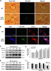
Fig. 1
Fig. 1. SiO2 exposure induces the EndMT in vivo and in vitro.
a Representative images of Sirius red staining in lung sections show the amount of Sirius red-stained collagen associated that co-localized with Tie2-GFP in mice treated with SiO2. b Representative images of immunohistochemical staining show the co-localization of TEK-GFP with α-SMA/Acta2 in TEK-GFP 287 Sato/JNju mice treated with SiO2, indicating that SiO2 induced the EndMT. c Representative western blot showing the effects of SiO2 (50 μg/cm2) on levels of the mesenchymal markers Col I/COL1A1, Col III/COL3A1 and α-SMA/Acta2. d Densitometric analyses of five separate experiments show that SiO2 increases Col I/COL1A1, Col III/COL3A1 and α-SMA/Acta2 expression in a time-dependent manner. *P < 0.05 vs. the expression of the corresponding protein at 0 h. e Representative western blot showing the effects of SiO2 (50 μg/cm2) on levels of the endothelial markers VE-Cad/Cdh-5 and PECAM 1/CD31. f Densitometric analyses of five separate experiments show that SiO2 decreases VE-Cad/Cdh-5 and PECAM 1/CD31 expression in a time-dependent manner. *P < 0.05 vs. corresponding protein at 0 h
Fig. 2
Fig. 2. SiO2 induces the proliferation and migration of MML1 cells.
a Representative images of immunocytochemical staining show that SiO2 (50 μg/cm2) decreases PECAM 1/CD31 expression and increases α-SMA/Acta2 expression in MML1 cells. b MTT assay showing the SiO2-induced increase in the viability of MML1 cells. *P < 0.05 vs. the 0-h group, n = 5. c Representative images showing the effects of SiO2 on GFP-labeled MML1 cell migration in scratch assays. Scale bar = 80 µm. d Quantification of the scratch gap distances in six separate experiments. *P < 0.05 vs. the control group at the corresponding time point
Fig. 3
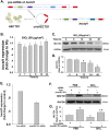
Fig. 3. SiO2 induces circHECTD1 expression in MML1 cells.
a Divergent primers amplified circRNAs from cDNAs, but not from gDNA. b As shown in the qRT-PCR analysis, circHECTD1 expression increased in the MML1 cells exposed to SiO2 (n = 5). *P < 0.05 vs. circHECTD1 expression at 0 h. c FISH analysis showing that circHECTD1 expression increases in MML1 cells exposed to SiO2. circHECTD1 was labeled with fluorescein isothiocyanate
Fig. 4
Fig. 4. circHECTD1 mediates the SiO2-induced EndMT.
a Representative western blot showing the effect of specific knockdown of circHECTD1 with siRNAs on SiO2-induced changes in endothelial and mesenchymal marker expression. The small pane shows the efficacy of circHECTD1-siRNA in reducing circHECTD1 expression. *P < 0.05 vs. the control group, n = 5. b Densitometric analyses of five separate experiments suggest that the SiO2-induced changes in the levels of endothelial and mesenchymal markers were attenuated by the circHECTD1-siRNA. *P < 0.05 vs. the level of the corresponding protein in the control group; #P < 0.05 vs. the level of the corresponding protein in the SiO2 group. c Representative images of immunocytochemical staining show that SiO2-induced α-SMA/Acta2 expression was attenuated by circHECTD1-siRNA
Fig. 5
Fig. 5. circHECTD1 is involved in SiO2-induced MML1 cell proliferation and migration.
a MTT assay showing that circHECTD1-siRNA attenuates the SiO2-induced increase in MML1 cell viability. *P < 0.05 vs. the control group; #P < 0.05 vs. the SiO2 group, n = 5. b Representative images show that the effect of SiO2 on GFP-labeled MML1 cell migration in scratch assays was attenuated by circHECTD1-siRNA. Scale bar = 80 µm. c Quantification of the scratch gap distances in six separate experiments. *P < 0.05 vs. the control group; #P < 0.05 vs. the SiO2 group
Fig. 6
Fig. 6. circHECTD1 is involved in SiO2-induced HECTD1 downregulation.
a In the SiO2-induced EndMT, circHECTD1 may regulate the expression of its host gene, hectd1. b As shown in the qRT-PCR analysis, levels of the hectd1 mRNA did not change in MML1 cells exposed to SiO2 (n = 5). c Representative western blot showing the effect of SiO2 on the level of the HECTD1 protein. d Densitometric analyses of five separate experiments suggest that SiO2 increases the level of the HECTD1 protein in MML1 cells. *P < 0.05 vs. protein expression at 0 h. e As shown in the qRT-PCR analysis, the expression of the hectd1 mRNA decreased in MML1 cells treated with circHECTD1-siRNA. *P < 0.05 vs. the control group, n = 5. f Representative western blot showing the effect of specific knockdown of circHECTD1 with siRNAs on SiO2-induced HECTD1 expression. g Densitometric analyses of five separate experiments suggest that SiO2-induced changes in HECTD1 expression are attenuated by circHECTD1-siRNA. *P < 0.05 vs. the levels of the corresponding protein in the control group; #P < 0.05 vs. the levels of the corresponding protein in the SiO2 group
Fig. 7
Fig. 7. HECTD1 is involved in the SiO2-induced EndMT in MML1 cells.
a Representative western blot and densitometric analyses showing the efficacy of HECTD1 CRISPR ACT at increasing the level of the HECTD1 protein. *P < 0.05 vs. the control group, n = 5. b Representative western blot showing the effect of specific upregulation of HECTD1 expression with ACT on SiO2-induced endothelial marker expression. c Densitometric analyses of five separate experiments suggest that the SiO2-induced changes in VE-Cad/Cdh-5 levels, but not PECAM 1/CD31 levels, are attenuated by HECTD1 ACT. *P < 0.05 vs. the level of the corresponding protein in the control group; #P < 0.05 vs. the level of the corresponding protein in the SiO2 group. d Representative western blot showing the effect of specific upregulation of HECTD1 with ACT on SiO2-induced mesenchymal marker expression. e Densitometric analyses of five separate experiments suggest that the SiO2-induced changes in mesenchymal markers are attenuated by HECTD1 ACT. *P < 0.05 vs. the level of the corresponding protein in the control group; #P < 0.05 vs. the level of the corresponding protein in the SiO2 group. f Representative images of immunocytochemical staining show that SiO2-induced α-SMA/Acta2 expression was attenuated by HECTD1 ACT
Fig. 8
Fig. 8. HECTD1 is involved in SiO2-induced MML1 cell proliferation and migration.
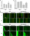
a MTT assay showing that HECTD1 ACT attenuated the SiO2-induced increase in MML1 cell viability. *P < 0.05 vs. the control group; #P < 0.05 vs. the SiO2 group, n = 5. b Representative images show that the effect of SiO2 on GFP-labeled MML1 cell migration in scratch assays was attenuated by HECTD1 ACT. Scale bar = 80 µm. c Quantification of the scratch gap distances in six separate experiments. *P < 0.05 vs. the control group; #P < 0.05 vs. the SiO2 group. d Representative images show that the effect of SiO2 on GFP-labeled MML1 cell migration in the nested matrix cell migration assay was attenuated by HECTD1 ACT. Scale bar = 80 µm
Fig. 9
Fig. 9. HECTD1 is involved in the EndMT in vivo.
a Representative images of immunohistochemical staining show that Tie2-GFP co-localization with HECTD1 decreased in TEK-GFP 287 Sato/JNju mice treated with SiO2. b Representative images of immunohistochemical staining show the expression patterns of PECAM 1/CD31 and HECTD1 in lung sections from healthy donors and patients with silicosis
Fig. 10
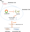
Fig. 10. Schematic diagram showing the mechanisms by which circHECTD1/HECTD1 regulate the SiO2-induced EndMT.
circHECTD1 expression is increased in endothelial cells exposed to SiO2. The increased expression leads to a subsequent decrease in HECTD1 expression, which, in turn, increases the levels of mesenchymal markers and decreases the levels of endothelial markers. Endothelial cell proliferation and migration are triggered, thereby contributing to irreversible fibrosis in patients with silicosis

Similar articles

Cited by

References

    1. Chao J, et al. Role of MCPIP1 in the endothelial–mesenchymal transition induced by silica. Cell. Physiol. Biochem. 2016;40:309–325. doi: 10.1159/000452547. - DOI - PubMed
    1. Leung CC, Yu IT, Chen W. Silicosis. Lancet. 2012;379:2008–2018. doi: 10.1016/S0140-6736(12)60235-9. - DOI - PubMed
    1. Arciniegas E, Neves CY, Carrillo LM, Zambrano EA, Ramirez R. Endothelial-mesenchymal transition occurs during embryonic pulmonary artery development. Endothelium. 2005;12:193–200. doi: 10.1080/10623320500227283. - DOI - PubMed
    1. Hashimoto N, et al. Endothelial–mesenchymal transition in bleomycin-induced pulmonary fibrosis. Am. J. Respir. Cell Mol. Biol. 2010;43:161–172. doi: 10.1165/rcmb.2009-0031OC. - DOI - PMC - PubMed
    1. Chen X, et al. Protective effect of spironolactone on endothelial-to-mesenchymal transition in HUVECs via Notch pathway. Cell. Physiol. Biochem. 2015;36:191–200. doi: 10.1159/000374063. - DOI - PubMed

Publication types

MeSH terms

LinkOut - more resources