. 2018 Mar 13;7(3):30.
doi: 10.1038/s41389-018-0039-5.

Expression of lncRNA MIR222HG co-transcribed from the miR-221/222 gene promoter facilitates the development of castration-resistant prostate cancer

Affiliations

Expression of lncRNA MIR222HG co-transcribed from the miR-221/222 gene promoter facilitates the development of castration-resistant prostate cancer

Tong Sun et al. Oncogenesis. .

Abstract

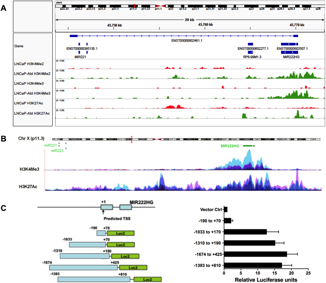
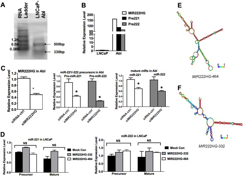
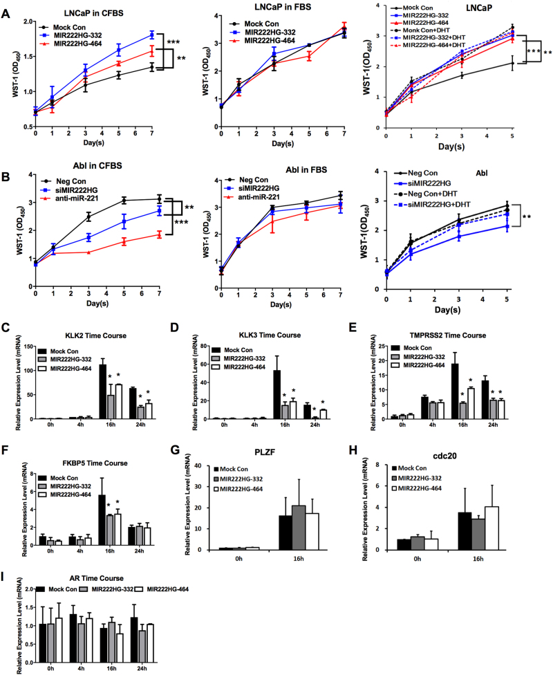
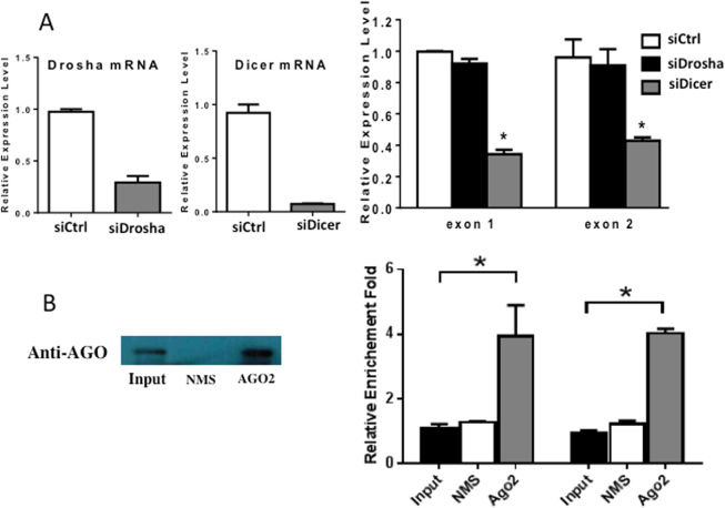
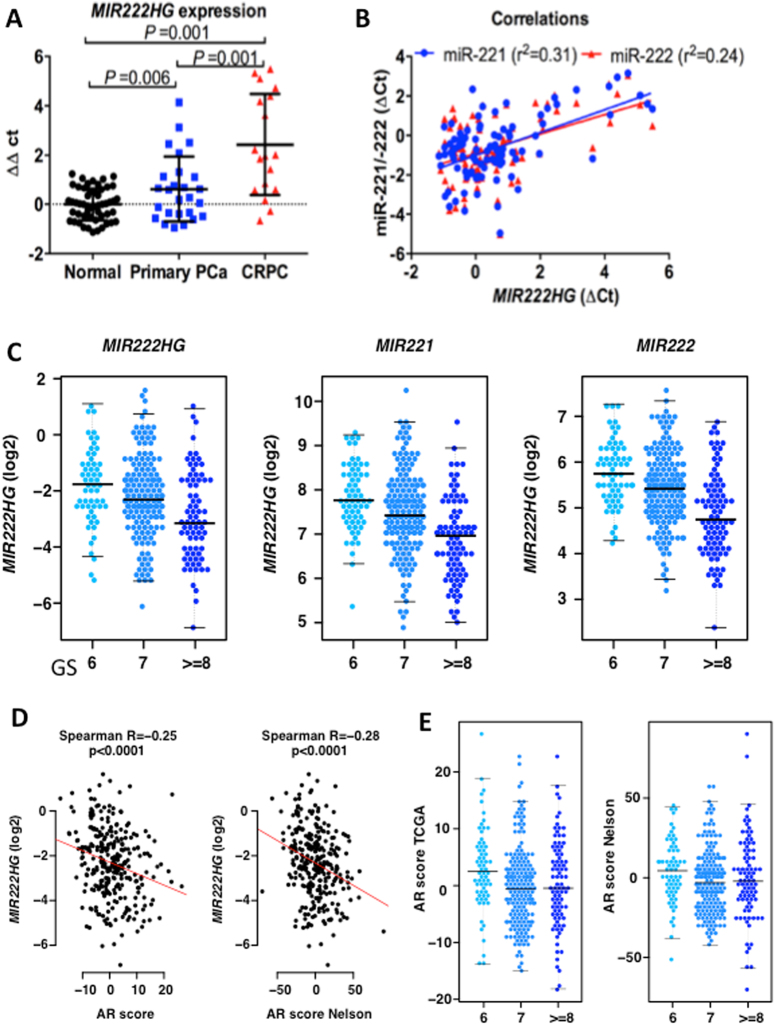
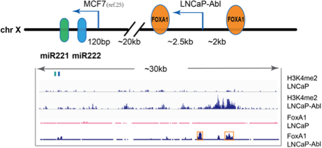
Mechanisms by which non-coding RNAs contribute to the progression of hormone-sensitive prostate cancer (PCa) (HSPC) to castration-resistant PCa (CRPC) remain largely unknown. We previously showed that microRNA-221/222 is up-regulated in CRPC and plays a critical role in modulating androgen receptor function during CRPC development. With further investigation, we characterized a putative promoter region located 23.3 kb upstream of the miR-221/222 gene, and this promoter is differentially activated in CRPC LNCaP-Abl cells, leading to the up-regulation of miR-221/222. Upon promoter activation, a set of polyadenylated long non-coding RNA (lncRNA) MIR222HGs was transcribed from this promoter region. Over-expression of these MIR222HGs increased androgen-independent cell growth and repressed the expression of androgen receptor-regulated dihydrotestosterone (DHT)-induced KLK3, TMPRSS2, and FKBP5 in HSPC LNCaP cells, hallmarks of the CRPC phenotype. Clinically, increased expression of MIR222HG is associated with PCa progression to CRPC. In primary tumors, expression levels of MIR222HG and miR-221/222 inversely correlate with Gleason score and androgen receptor (AR) pathway activity. Interestingly, MIR222HG is Argonaute 2-bound and its expression is Dicer 1-dependent, suggesting its functional association with the RNA-induced silencing complex. Further studies led to the hypothesis that MIR222HG may potentially affect miR-mediated expression silencing, subsequently leading to AR reprogramming. Our study highlights an essential role of a non-coding RNA in CRPC development and that differential activation of a single promoter can up-regulate two different types of non-coding RNAs, miR-221/222 and lncRNA MIR222HG, in CRPC. Additionally, this study reveals a novel function of lncRNAs as a modulator of Argonaute-mediated RNA-induced silencing complex.


Conflict of interest statement

The authors declare that they have no conflict of interest.

Figures

Fig. 1
Fig. 1. Identification of a promoter driving the transcription of the miR-221/222 locus.
a H3K4Me3, H3K4Me2, and H3K27Ac marks in LNCaP and LNCaP-Abl. Top panel, the diagram of miR-221/222 gene and MIR222HG loci in Chr. Xp11. 3. All CHIP-seq data were documented in www.cistrome.org. b Upper panel, H3K4Me3 marks on seven cell lines from ENCODE hg19; lower panel, H3K27Ac marks on seven non-PCa cell lines (GM12878, Hi-hESC, HSMM, HUVEC, K562, NHEK, and NHLF) from ENCODE hg19 (www.genome.ucsc.edu). c Characterization of a promoter region at the 5′ region of the MIR222HG gene. Left panel, fragments of the promoter region were cloned into the upstream of a luciferase reporter gene. Right panel, promoter luciferase reporter assays in LNCaP-Abl cells. Relative luciferase activities were calculated in relative to the vector control (Renilla luciferase vector without a promoter) whose activity was arbitrarily set as 1
Fig. 2
Fig. 2. Characterization of MIR222HG lncRNA.
a Northern blot of MIR222HG lncRNA in LNCaP-Abl cells. Poly(A)-tailed RNA from LNCaP-Abl was separated on a 1.5% denaturing agarose gel and was hybridized with DIG-labeled DNA probes. Two horizontal arrows indicate two different isoforms of MIR222HG lncRNA. b The expression levels of MIR222HG (white bars), miR-221 precursors (black bars), and miR-222 precursors (gray bars) in LNCaP and LNCaP-Abl cell lines. c Knocking down MIR222HG lncRNA reduced the expression levels of precursors and mature miR-221/222. Left panel, MIR222HG siRNA efficiency in LNCaP-Abl cells; middle panel, miR-221/222 precursors in LNCaP-Abl cells after MIR222HG knocking down; right panel, mature miR-221/222 expression in LNCaP-Abl after MIR222HG knocking down. d Over-expressing MIR222HG lncRNA had no impact on miR-221/222 expression. Left panel, the level of precursor and mature miR-221 in LNCaP after over-expressing two different isoforms of MIR222HG in LNCaP cells; right panel, the level of precursor and mature miR-222 in LNCaP after over-expressing two different isoforms of MIR222HG. e, f RNAfold secondary structure predictions of MIR222HG-464 and MIR222HG-332, with the minimum free energy as −108.7l and −82.7 kcal/mol, respectively. The color scale indicates high (red) to low (blue) probabilities of base pairing. RNA secondary structures and energies were predicted using RNAfold from the Vienna RNA package
Fig. 3
Fig. 3. The impact of MIR222HG expression levels on CRPC characteristics.
a Effect of MIR222HG expression level on the growth of LNCaP. LNCaP cells that were transfected with the empty vector (Mock Con, black lines), MIR222HG-332 bp isoform (blue lines) or 464 bp isoform (red lines) in hormone-free medium (CFBS, left panel), in regular medium (FBS, middle panel), or in CFBS plus 10 nM DHT (right panel; solid lines indicating CFBS only; dotted lines indicating CFBS + DHT). b Effect of MIR222HG expression level on the growth of LNCaP-Abl. LNCaP-Abl cells that were transfected with the negative siRNA control (black lines), anti-miR-221 (red lines), or MIR222HG siRNAs (blue lines) and kept in hormone-free medium (CFBS, left panel), in regular medium (FBS, middle panel), or in CFBS treated with 10 nM DHT(right panel; solid lines indicating CFBS only; dotted lines indicating CFBS + DHT). Cell growth was measured by the WST-1 assay. Triplicate experiments were performed for each set. The data represents mean ± S.D. (n = 3). *The WST-1 fold changes at day 7 after transfection with a p value <0.01 (**) or p value <0.001 (***) in one-way ANOVA. c–h The impact of MIR222HG expression level on the AR-mediated transcription in response to the DHT treatment. Quantitative analysis of the expression level of KLK2 (c), KLK3 (d), TMPRSS2 (e), FKBP5 (f), PLZF (g), and cdc20 (h) in LNCaP upon DHT treatment. i The AR mRNA expression level in MIR222HG-over-expressing cells. The relative expression levels of AR and AR-mediated genes in each sample were normalized with the expression level of GAPDH. Values represent the fold differences relative to those in cells without any drug treatment or transfection (Mock), which were set as 1.0. *The fold changes of those transfected samples compared with their corresponding negative controls show a p value <0.05 in one-way ANOVA
Fig. 4
Fig. 4. Relationship of MIR222HGs with Drosha, Dicer, and Argonaute 2 (AGO2).
a The impact of knocking down Drosha or Dicer 1 on the expression level of MIR222HGs. LNCaP-Abl was transfected with siDrosha, siDicer 1, and siCtrl (siControl), separately. Total RNAs were isolated post-transfection and RT-PCRs were performed to determine the mRNA levels of Drosha and Dicer 1 in the left panel and the level of MIR222HGs as measured by primers specific for exon 1 and exon 2, in the right panel. b The association of MIR222HGs with AGO2. Total cell lysates of LNCaP-Abl were IPed with non-immune IgG and anti-AGO2 antibody, respectively. The Ago 2 protein level in the input and IP pull-down materials were measured by western blot as shown in the left panel. The amount of MIR222HGs in IP pull-down materials was analyzed by RT-PCR using probes specific for exon 1 and exon 2, respectively, as shown in the right-hand panel
Fig. 5
Fig. 5. MIR222HG expression pattern in normal prostate, primary tumors, and CRPC.
a Comparison of the MIR222HG expression. Expression levels of MIR222HG were measured by quantitative RT-PCR on 86 normal prostate tissues (black dots), 34 hormone-sensitive primary prostate tumors (blue squares), and 17 metastatic CRPC tissues (red triangles). All expression levels were normalized by 28S ribosome RNA (RPS28). Values represent in qPCR cycle differences relative to the mean expression level of normal tissue, which was set as 0. P values were estimated by one-way ANOVA. b The expression correlation between MIR222HG and miR-221 or miR-222 in human prostate tissue specimen. The Pearson's correlation coefficient indexes (r2) were calculated by comparing normalized qPCR cycle values (∆cts) in detecting miR-221 (blue) or miR-222 (red) with ∆ct value of MIR222HG. c Association of expression levels of MIR222HG, miR-221, and miR-222 with GS in primary tumors of the TCGA data set (p < 0.0001 Kruskal–Wallis test). d Correlation of MIR222HG expression level with AR activity (indicated by AR score) in primary tumors of the TCGA data set. AR activity score was inferred by the induction of AR target genes,. AR score Nelson indicates that the AR score was re-evaluated using the AR gene index previously published by Dr. Nelson’s laboratory. e Correlation of GS with AR activity score
Fig. 6
Fig. 6. A graphical representation of the miR-221/222 locus.
Upper panel demonstrates a schematic diagram of miR-221/222 genomic loci. The proximal promoter region was initially identified in MCF7 and the distal promoter region was identified in LNCaP-Abl as described in Fig. 1. Lower panel demonstrates the ChIP-seq enrichment of H3K4Me2 and FoxA1 binding sites in LNCaP and LNCaP-Abl cells. Each data track shown is on the same scale for both LNCaP and LNCaP-Abl cells

Similar articles

Cited by

References

    1. Siegel RL, Miller KD, Jemal A. Cancer statistics, 2015. CA Cancer J. Clin. 2015;65:5–29. doi: 10.3322/caac.21254. - DOI - PubMed
    1. Heinlein CA, Chang C. Androgen receptor in PCa. Endocr. Rev. 2004;25:276–308. doi: 10.1210/er.2002-0032. - DOI - PubMed
    1. Harris WP, Mostaghel EA, Nelson PS, Montgomery B. Androgen deprivation therapy: progress in understanding mechanisms of resistance and optimizing androgen depletion. Nat. Clin. Pract. Urol. 2009;6:76–85. doi: 10.1038/ncpuro1296. - DOI - PMC - PubMed
    1. Yuan X, et al. Androgen receptor functions in castration-resistant PCa and mechanisms of resistance to new agents targeting the androgen axis. Oncogene. 2014;33:2815–2825. doi: 10.1038/onc.2013.235. - DOI - PMC - PubMed
    1. Bluemn EG, Nelson PS. The androgen/androgen receptor axis in PCa. Curr. Opin. Oncol. 2012;24:251–257. doi: 10.1097/CCO.0b013e32835105b3. - DOI - PMC - PubMed