. 2018 Mar 14;9(3):397.
doi: 10.1038/s41419-018-0427-y.

Inhibition of Sec61-dependent translocation by mycolactone uncouples the integrated stress response from ER stress, driving cytotoxicity via translational activation of ATF4

Affiliations

Inhibition of Sec61-dependent translocation by mycolactone uncouples the integrated stress response from ER stress, driving cytotoxicity via translational activation of ATF4

Joy Ogbechi et al. Cell Death Dis. .

Abstract

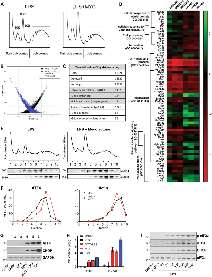
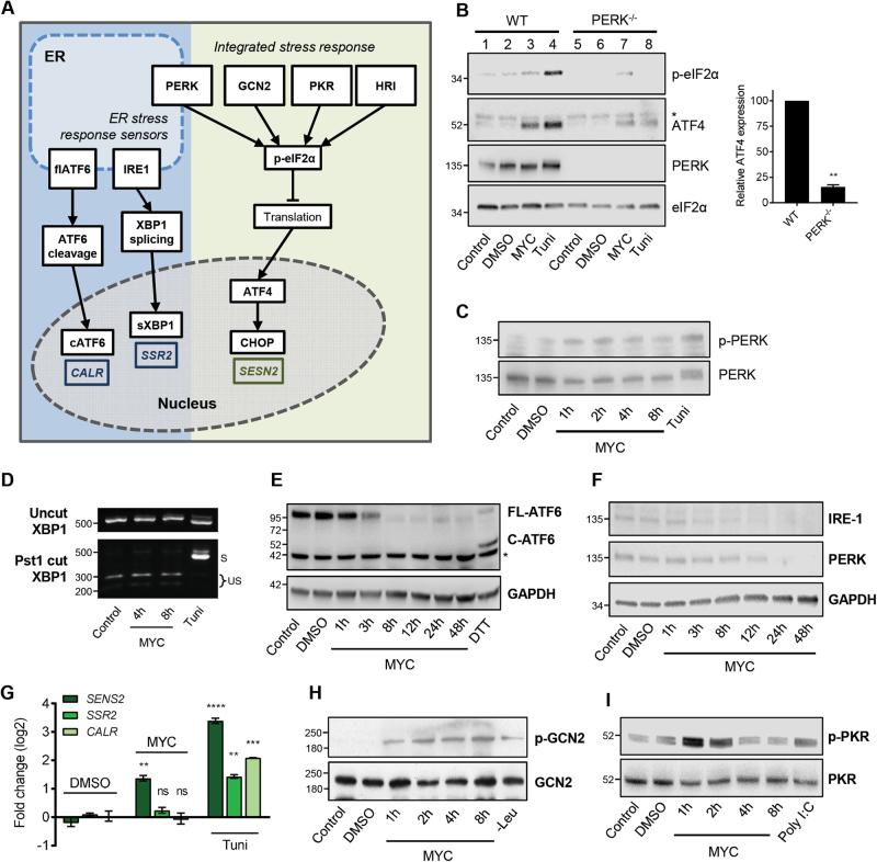
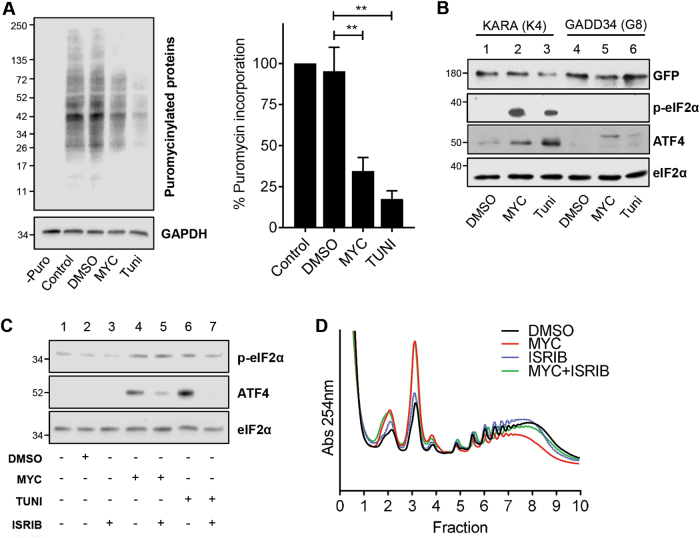
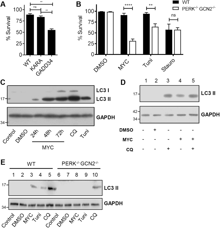
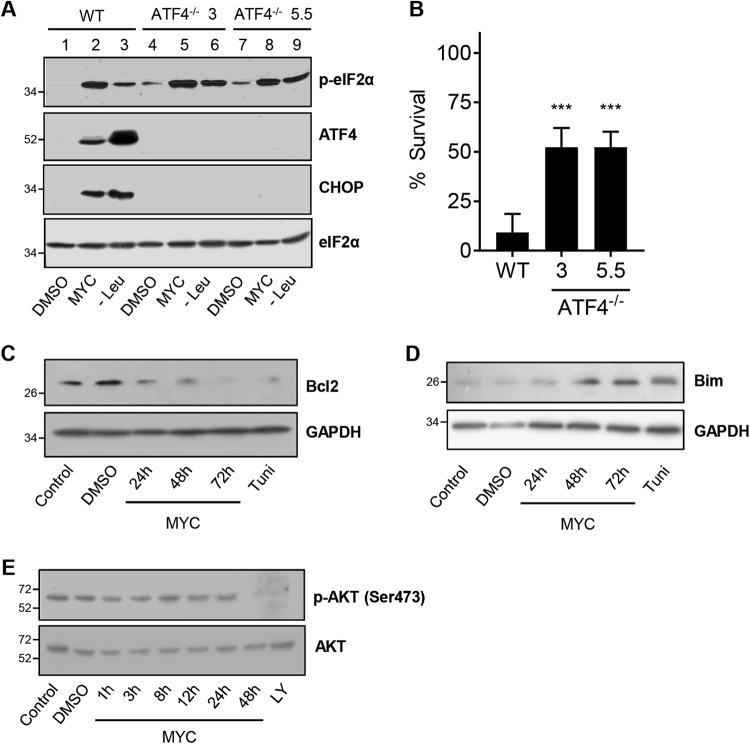
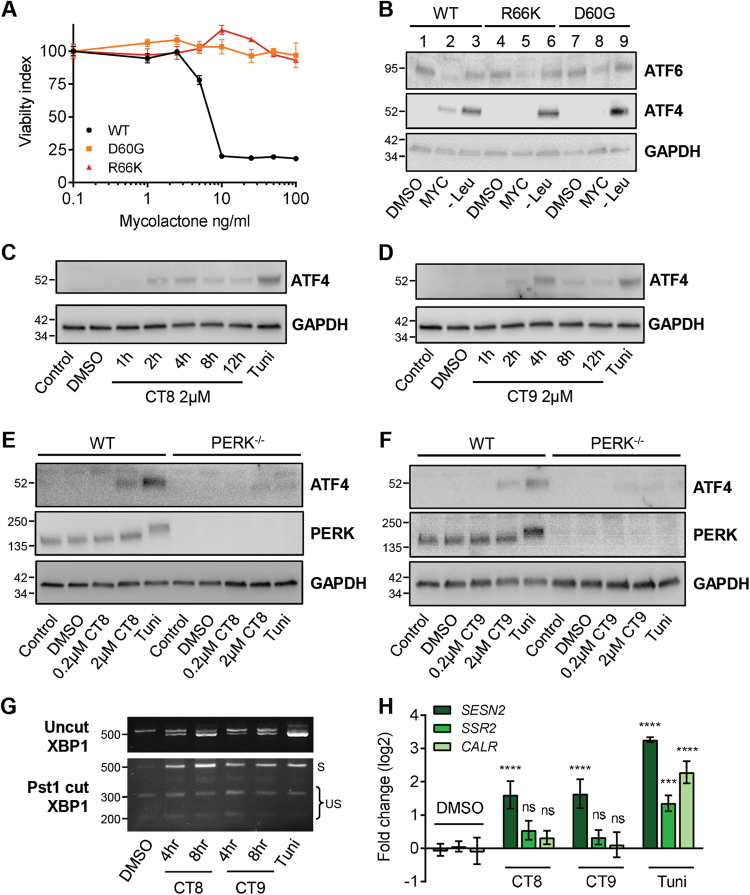
Mycolactone is the exotoxin virulence factor of Mycobacterium ulcerans that causes the neglected tropical disease Buruli ulcer. We recently showed it to be a broad spectrum inhibitor of Sec61-dependent co-translational translocation of proteins into the endoplasmic reticulum (ER). An outstanding question is the molecular pathway linking this to its known cytotoxicity. We have now used translational profiling to better understand the reprogramming that occurs in cells exposed to mycolactone. Gene ontology identified enrichment in genes involved in cellular response to stress, and apoptosis signalling among those showing enhanced translation. Validation of these results supports a mechanism by which mycolactone activates an integrated stress response meditated by phosphorylation of eIF2α via multiple kinases (PERK, GCN, PKR) without activation of the ER stress sensors IRE1 or ATF6. The response therefore uncouples the integrated stress response from ER stress, and features translational and transcriptional modes of genes expression that feature the key regulatory transcription factor ATF4. Emphasising the importance of this uncoupled response in cytotoxicity, downstream activation of this pathway is abolished in cells expressing mycolactone-resistant Sec61α variants. Using multiple genetic and biochemical approaches, we demonstrate that eIF2α phosphorylation is responsible for mycolactone-dependent translation attenuation, which initially protects cells from cell death. However, chronic activation without stress remediation enhances autophagy and apoptosis of cells by a pathway facilitated by ATF4 and CHOP. Our findings demonstrate that priming events at the ER can result in the sensing of stress within different cellular compartments.


Conflict of interest statement

The authors declare that they have no conflict of interest.

Figures

Fig. 1
Fig. 1. Translational microarray of cells exposed to mycolactone identifies ATF4 as translationally upregulated.
ag RAW264.7 cells pre-incubated for 1 h (mycolactone (MYC) or DMSO) then stimulated with 100 ng/ml LPS for 4 h or tunicamycin alone (Tuni). ae Cell lysates were subject to polysome profiling and translational microarrays, as described in the text. a Mycolactone induces changes in polysome profiles. RNA purified from subpolysomal fractions (1–5) and polysomal fractions (6–10) was pooled and used in microarray analysis as described in Methods. b Scatter plot for probes in the microarray. Black and blue dots represent probes enriched in either polysomes or sub-polysomes respectively. Rank product analysis rules out changes in transcription and achieves a high validation rate for translationally regulated targets. c Summary of microarray data following translational profiling analysis as described in Methods. d Heatmap showing representative data for genes in eight significantly overrepresented gene ontology groups (p < 0.05), identified by PANTHER. e Northern blotting for transcripts in individual gradient fractions from LPS stimulated RAW264.7 cells, the migration of 18S rRNA is indicated; quantified in (f); n = 3 independent experiments. g Cell lysates were analysed by immunoblotting. h Relative fold change (ΔΔCt) for steady-state mRNA levels determined by one-step qRT-PCR on total RNA (Mean ± SEM, n = 3 independent experiments). i HeLa cells were treated as shown and lysates were analysed by immunoblotting. All immunoblots show the approximate migration of molecular weight markers in kDa. See also Figs. S1 and S2, Tables S1 and S2
Fig. 2
Fig. 2. Mycolactone uncouples the integrated response from the unfolded protein response via a pathway that implicates multiple eIF2α kinases.
a Cartoon representation of the ISR and ER stress response sensors and consequences. Genes that are specifically induced by the three ER sensors are shown. b Wild-type (WT) or knockout MEFs were treated with either mycolactone (MYC), DMSO or tunicamycin (Tuni), and the lysates were analysed by immunoblotting. * indicates a cross-reactive band. Relative semi-quantified signal intensities for ATF4 (Mean ± SEM, n = 3 independent experiments). c, h, i HeLa cells were treated as shown and lysates were analysed by immunoblotting. Representative data from n > 3 independent experiments. d HeLa cells were treated as shown. Total RNA was isolated and used as a template for RT-PCR of XBP-1 (upper panel) which was then digested with Pst1 and separated on a 2% agarose gel (lower panel). The migration of molecular weight markers in bp is indicated. S spliced, US unspliced. e, f HeLa cells were treated with DMSO for 48 h, DTT for 1 h or mycolactone (MYC) for the indicated duration (up to 48 h). Equal protein quantities in lysates were analysed by immunoblotting. * indicates a cross-reactive band. g HeLa cells were treated as shown for 10 h. Relative fold change (ΔΔCt) for steady-state mRNA levels determined by one-step qRT-PCR on total RNA (Mean ± SEM of three independent experiments). All immunoblots show the approximate migration of molecular weight markers in kDa. See also Fig. S3
Fig. 3
Fig. 3. eIF2α phosphorylation and translational control drive ATF4 expression in cells exposed to mycolactone.
a Immunoblot analysis of newly synthesised puromycilated proteins prepared from HeLa cells exposed to DMSO, mycolactone (MYC) or tunicamycin (Tuni) for 12 h. Relative quantified signal intensities are shown (Mean ± SEM, n = 3 independent experiments). b HeLa-gs cells stably expressing GFP-GADD34 (clone 8) or GFP-KARA (clone 4), an inactive mutant of GADD34, were exposed for 5 h. Lysates were analysed by immunoblotting. c HeLa cells were pre-treated with ISRIB for 1 h, followed by exposure to mycolactone for 8 h. Lysates were analysed by immunoblotting. d RAW246.7 cells were exposed to either DMSO, mycolactone or 100 nM ISRIB for 5 h. For co-incubation cells were pre-treated with ISRIB for 1 h prior to addition of mycolactone. Cell lysates were subject to polysome profiling. All immunoblots show the approximate migration of molecular weight markers in kDa. All data representative of at least three independent experiments
Fig. 4
Fig. 4. The phosphorylation of eIF2α protects cells by a mechanism that involves adaptive autophagy.
a HeLa-gs cells stably expressing GFP-GADD34 (clone 8) or GFP-KARA (clone 4), an inactive mutant of GADD34, were treated with mycolactone for 4 days The number of apoptotic cells (positive for both active caspase 3/7 and PI) were determined for three fields and expressed as a proportion of total cells (Mean ± SEM n = 4 independent experiments). b Wild-type and PERK−/− GCN2−/− MEFs were treated with mycolactone for 24 h and analysed by confocal microscopy as in (a). c, d HeLa cells were treated as shown or with chloroquine (CQ) for 12 h. Equal protein quantities in lysates were analysed by immunoblotting. e WT and PERK/ GCN2−/− MEFs were treated as in (c). Lysates were analysed by immunoblotting. All data representative of at least three independent experiments. All immunoblots show the approximate migration of molecular weight markers in kDa. See also Fig. S4
Fig. 5
Fig. 5. ATF4 promotes mycolactone-mediated cytotoxicity.
a, b Wild-type HeLa cells and two different ATF4−/− clones (3 and 5.5) were treated with either DMSO, mycolactone (MYC) or starved of leucine (Leu). a Lysates from 24-h-treated cells were analysed by immunoblotting. b After 4 days the % survival of cells was determined by staining of cells with propidium iodide (PI), cell event (detects active caspase 3/7) and DRAQ5. The number of live cells (negative for both active caspase 3/7 and PI) in three fields was determined and expressed as a proportion of total cells (Mean ± SEM, n = 3 independent experiments). ce HeLa cells were treated as shown or with LY294002 (LY) for 1 h. Equal protein quantities in lysates were analysed by immunoblotting. All immunoblots show the approximate migration of molecular weight markers in kDa. See also Fig. S5
Fig. 6
Fig. 6. Uncoupling of the ISR from ER stress is a consequence of mycolactone’s effect on the Sec61 translocon, but is not a general feature of translocation inhibition.
a, b An unbiased screen for mycolactone-resistant clones was performed in HCT-116 cells, yielding clones with heterozygous missense mutations in Sec61A1. Parental HCT-116 cells and representative clones with D60G and R66K were analysed. a Normalised viability index of cells treated with mycolactone for 5 days, assessed by MTT assay. b Immunoblot of lysates that were treated with DMSO, mycolactone or starved of Leucine (Leu) for 24 h. c, d Wild-type MEFs were treated with CT8 (c) or CT9 (d) for the indicated times or tunicamycin (Tuni). Lysates were analysed by immunoblotting. e, f Wild-type (WT) or PERK−/− MEFs were treated as shown and the lysates were analysed by immunoblotting. g Wild-type MEFs were treated as shown. Total RNA was isolated and used as a template for RT-PCR of XBP-1 (upper panel) which was then digested with Pst1 and separated on a 2% agarose gel (lower panel). The migration of molecular weight markers in bp is indicated. S spliced, US unspliced. All data representative of at least two independent experiments. h HeLa cells were treated as shown for 10 h. Relative fold change (ΔΔCt) for steady-state mRNA levels (Mean ± SEM, n = 3 independent experiments). All immunoblots show the approximate migration of molecular weight markers in kDa

References

    1. George KM, et al. Mycolactone: a polyketide toxin from Mycobacterium ulcerans required for virulence. Science. 1999;283:854–857. doi: 10.1126/science.283.5403.854. - DOI - PubMed
    1. Walsh DS, Portaels F, Meyers WM. Buruli ulcer: advances in understanding Mycobacterium ulcerans infection. Dermatol. Clin. 2011;29:1–8. doi: 10.1016/j.det.2010.09.006. - DOI - PubMed
    1. Sarfo FS, Phillips R, Wansbrough-Jones M, Simmonds RE. Recent advances: role of mycolactone in the pathogenesis and monitoring of Mycobacterium ulcerans infection/Buruli ulcer disease. Cell. Microbiol. 2016;18:17–29. doi: 10.1111/cmi.12547. - DOI - PMC - PubMed
    1. Hall BS, et al. The pathogenic mechanism of the Mycobacterium ulcerans virulence factor, mycolactone, depends on blockade of protein translocation into the ER. PLoS Pathog. 2014;10:e1004061. doi: 10.1371/journal.ppat.1004061. - DOI - PMC - PubMed
    1. McKenna M, Simmonds RE, High S. Mechanistic insights into the inhibition of Sec61-dependent co- and post-translational translocation by mycolactone. J. Cell Sci. 2016;129:1404–1405. doi: 10.1242/jcs.182352. - DOI - PMC - PubMed

Publication types

MeSH terms