. 2018 Mar 14;9(1):1069.
doi: 10.1038/s41467-018-03369-8.

Circular DNA elements of chromosomal origin are common in healthy human somatic tissue

Affiliations

Circular DNA elements of chromosomal origin are common in healthy human somatic tissue

Henrik Devitt Møller et al. Nat Commun. .

Abstract

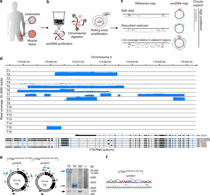
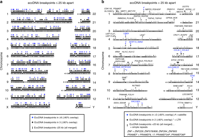
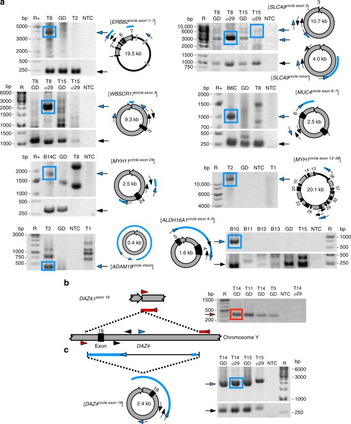
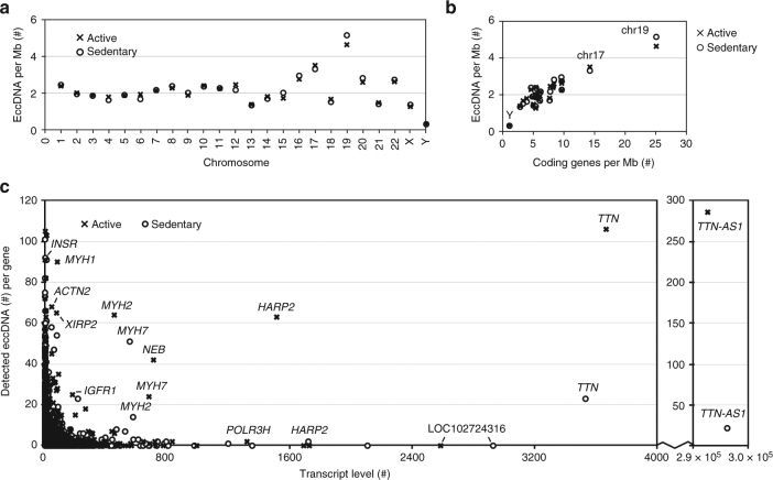
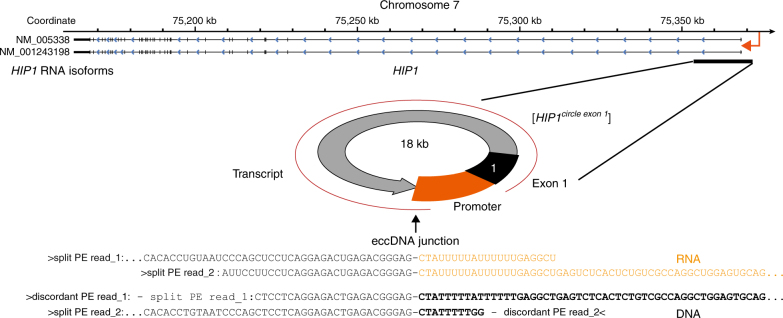
The human genome is generally organized into stable chromosomes, and only tumor cells are known to accumulate kilobase (kb)-sized extrachromosomal circular DNA elements (eccDNAs). However, it must be expected that kb eccDNAs exist in normal cells as a result of mutations. Here, we purify and sequence eccDNAs from muscle and blood samples from 16 healthy men, detecting ~100,000 unique eccDNA types from 16 million nuclei. Half of these structures carry genes or gene fragments and the majority are smaller than 25 kb. Transcription from eccDNAs suggests that eccDNAs reside in nuclei and recurrence of certain eccDNAs in several individuals implies DNA circularization hotspots. Gene-rich chromosomes contribute to more eccDNAs per megabase and the most transcribed protein-coding gene in muscle, TTN (titin), provides the most eccDNAs per gene. Thus, somatic genomes are rich in chromosome-derived eccDNAs that may influence phenotypes through altered gene copy numbers and transcription of full-length or truncated genes.


Conflict of interest statement

The authors declare no competing interests.

Figures

Fig. 1
Fig. 1
Circle-Seq method for mapping of eccDNA. a Leukocyte and muscle samples from 16 healthy subjects (T1–T16, n = 16). b Purification of eccDNA through column separation, exonuclease treatment, and rolling-circle amplification. c Detection of eccDNA based on structural-read variants and coverage (soft-clipped, split, red; concordant, gray; and discordant reads, blue). d Read coverage display (log-scale) at the titin gene, TTN, from muscle samples. e EccDNA from T5 and T6 illustrated (black boxes, exons), outward PCR validation (blue arrows), inward PCR (black arrows), and gel-image of T5 and T6 PCR products next to controls: GD, human genomic DNA; φ, phi29-amplified eccDNA sample without detected [TTNcircle]. f Sequence of [TTNcircle exon 44-52] T6 PCR product at junction
Fig. 2
Fig. 2
EccDNA sizes, read distribution on plasmids and repetitive elements. Number of eccDNAs relative to size in kb for 1 × 106 muscle nuclei from each individual. a Physically inactive men (n = 8); b active men (n = 8). EccDNA kb sizes are in bins of 0.1 log-fold intervals. c Percent mapped reads per sample for plasmids added to muscle samples at indicated copy numbers (median, white circles). Repetitive regions from total mapped reads for each muscle-derived eccDNA sample (T1–T16); median, white circles (n = 16)
Fig. 3
Fig. 3
Genomic distribution of eccDNA breakpoints. Distribution of detected chromosomal breakpoints across the human genome with read support for circular DNA, in sizes of ≤25 kb (total 147,186) and b >25 kb apart (total 1,014) in muscle (T1–T16, n = 16) and leukocyte (B1–B16, n = 16) samples. EccDNA detected once, black lines; eccDNA detected repeatedly in several individuals with ≥90% overlap, circles
Fig. 4
Fig. 4
EccDNA validation and DNA deletion. a Gel images for a validated subset (n = 9) of eccDNAs by outward PCR (blue arrows), inward PCR (black arrows), gel electrophoresis, and Sanger sequencing. EccDNAs are named according to gene content; black boxes, exons. Template: T1–T16, muscle B1–B16 leukocyte, phi29 (φ) amplified eccDNA, GD genomic DNA, NTC nontemplate control. Sequenced PCR products are in boxes and alignment of resultant sequence are red and blue lines. b Confirmed verification of DAZ4exon 18 deletion and c [DAZ4circle exon 18] at identical coordinates within the DAZ4 locus by inward and outward PCR (oligos arrows, inward red, outward, blue)
Fig. 5
Fig. 5
EccDNA frequency relative to chromosome, gene density and transcription. Muscle-derived eccDNA counts per Mb from physically active (x, n = 8) and inactive men (o, n = 8) per: a chromosome and b coding genes per Mb (chromosome Y, 17 and 19 are marked). c EccDNA counts per gene relative to average transcription level of muscle sample (two sets, each group n = 8)
Fig. 6
Fig. 6
EccDNA transcript from the [HIP1circle exon 1]. Display of chromosome 7 at the HIP1 gene and detected [HIP1circle exon 1]. The two sequenced RNA reads (black and orange text) from participant T8 overlap perfectly to the two sequenced DNA reads from participant T8 (black and bold text font)

Similar articles

Cited by

References

    1. Parshad R, Sanford KK, Jones GM. Chromosomal radiosensitivity during the G2 cell-cycle period of skin fibroblasts from individuals with familial cancer. Proc. Natl Acad. Sci. USA. 1985;82:5400–5403. doi: 10.1073/pnas.82.16.5400. - DOI - PMC - PubMed
    1. Wright JA, et al. DNA amplification is rare in normal human cells. Proc. Natl Acad. Sci. USA. 1990;87:1791–1795. doi: 10.1073/pnas.87.5.1791. - DOI - PMC - PubMed
    1. Iafrate AJ, et al. Detection of large-scale variation in the human genome. Nat. Genet. 2004;36:949–951. doi: 10.1038/ng1416. - DOI - PubMed
    1. Redon R, et al. Global variation in copy number in the human genome. Nature. 2006;444:444–454. doi: 10.1038/nature05329. - DOI - PMC - PubMed
    1. Besenbacher S, et al. Novel variation and de novo mutation rates in population-wide de novo assembled Danish trios. Nat. Commun. 2015;6:5969. doi: 10.1038/ncomms6969. - DOI - PMC - PubMed

Publication types