

Bone marrow mesenchymal stem cells protect against n-hexane-induced neuropathy through beclin 1-independent inhibition of autophagy
- PMID: 29540747
- PMCID: PMC5852116
- DOI: 10.1038/s41598-018-22857-x
Bone marrow mesenchymal stem cells protect against n-hexane-induced neuropathy through beclin 1-independent inhibition of autophagy
Abstract
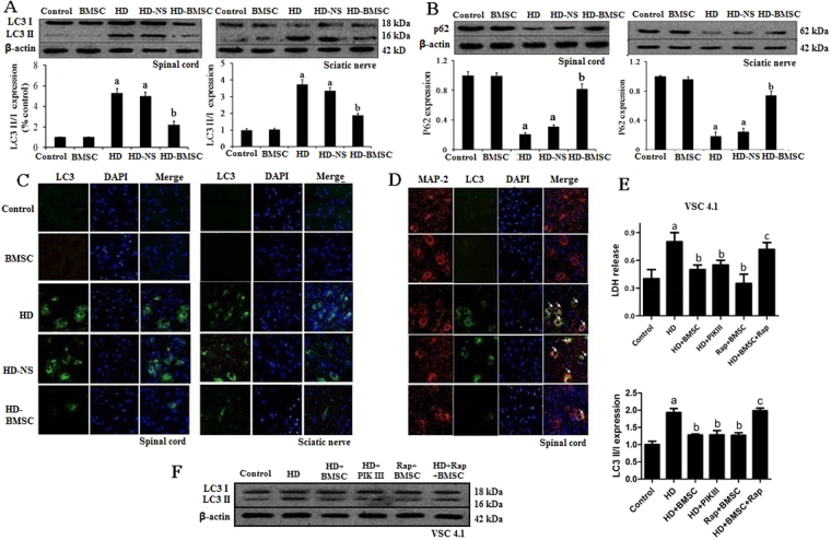
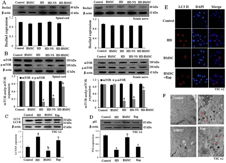
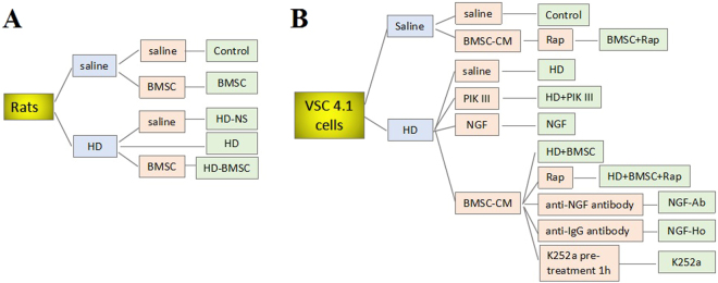
Chronic exposure to n-hexane, a widely used organic solvent in industry, induces central-peripheral neuropathy, which is mediated by its active metabolite, 2,5-hexanedione (HD). We recently reported that transplantation of bone marrow-mesenchymal stem cells (BMSC) significantly ameliorated HD-induced neuronal damage and motor deficits in rats. However, the mechanisms remain unclear. Here, we reported that inhibition of HD-induced autophagy contributed to BMSC-afforded protection. BMSC transplantation significantly reduced the levels of microtubule-associated protein 1 light chain 3-II (LC3-II) and the degradation of sequestosome-1 (p62) in the spinal cord and sciatic nerve of HD-intoxicated rats. Downregulation of autophagy by BMSC was also confirmed in VSC4.1 cells exposed to HD. Moreover, inhibition of autophagy by PIK III mitigated the neurotoxic effects of HD and, meanwhile, abolished BMSC-afforded neuroprotection. Furthermore, we found that BMSC failed to interfere with Beclin 1, but promoted activation of mammalian target of rapamycin (mTOR). Unc-like kinse 1 (ULK1) was further recognized as the downstream target of mTOR responsible for BMSC-mediated inhibition of autophagy. Altogether, BMSC transplantation potently ameliorated HD-induced autophagy through beclin 1-independent activation of mTOR pathway, providing a novel insight for the therapeutic effects of BMSC against n-hexane and other environmental toxicants-induced neurotoxicity.
Conflict of interest statement
The authors declare no competing interests.
Figures


Similar articles
-
Bone marrow mesenchymal stem cells conditioned medium protects VSC4.1 cells against 2,5-hexanedione-induced autophagy via NGF-PI3K/Akt/mTOR signaling pathway.Brain Res. 2018 Oct 1;1696:1-9. doi: 10.1016/j.brainres.2018.04.028. Epub 2018 Apr 27. Brain Res. 2018. PMID: 29705604
-
Bone marrow mesenchymal stem cells attenuate 2,5-hexanedione-induced neuronal apoptosis through a NGF/AKT-dependent pathway.Sci Rep. 2016 Oct 5;6:34715. doi: 10.1038/srep34715. Sci Rep. 2016. PMID: 27703213 Free PMC article.
-
Bone marrow mesenchymal stem cells promote remyelination in spinal cord by driving oligodendrocyte progenitor cell differentiation via TNFα/RelB-Hes1 pathway: a rat model study of 2,5-hexanedione-induced neurotoxicity.Stem Cell Res Ther. 2021 Aug 4;12(1):436. doi: 10.1186/s13287-021-02518-z. Stem Cell Res Ther. 2021. PMID: 34348774 Free PMC article.
-
NGF mediates protection of mesenchymal stem cells-conditioned medium against 2,5-hexanedione-induced apoptosis of VSC4.1 cells via Akt/Bad pathway.Mol Cell Biochem. 2020 Jun;469(1-2):53-64. doi: 10.1007/s11010-020-03727-5. Epub 2020 Apr 11. Mol Cell Biochem. 2020. PMID: 32279149
-
The functional mechanism of bone marrow-derived mesenchymal stem cells in the treatment of animal models with Alzheimer's disease: crosstalk between autophagy and apoptosis.Stem Cell Res Ther. 2022 Mar 3;13(1):90. doi: 10.1186/s13287-022-02765-8. Stem Cell Res Ther. 2022. PMID: 35241159 Free PMC article. Review.
Cited by
-
Human Bone Marrow Mesenchymal Stem Cells-Derived Exosomal miRNA-21-5p Inhibits Lidocaine-Induced Apoptosis in SH-SY5Y Neuroblastoma Cells.Iran J Public Health. 2023 Apr;52(4):756-765. doi: 10.18502/ijph.v52i4.12446. Iran J Public Health. 2023. PMID: 37551179 Free PMC article.
-
Autophagy inhibition via Becn1 downregulation improves the mesenchymal stem cells antifibrotic potential in experimental liver fibrosis.J Cell Physiol. 2020 Mar;235(3):2722-2737. doi: 10.1002/jcp.29176. Epub 2019 Sep 11. J Cell Physiol. 2020. PMID: 31508820 Free PMC article.
-
Targeting autophagy to overcome drug resistance: further developments.J Hematol Oncol. 2020 Nov 25;13(1):159. doi: 10.1186/s13045-020-01000-2. J Hematol Oncol. 2020. PMID: 33239065 Free PMC article. Review.
-
Mesenchymal stem cells secretome: The cornerstone of cell-free regenerative medicine.World J Stem Cells. 2020 Dec 26;12(12):1529-1552. doi: 10.4252/wjsc.v12.i12.1529. World J Stem Cells. 2020. PMID: 33505599 Free PMC article. Review.
-
2,5-Hexanedione mediates neuronal apoptosis through suppression of NGF via PI3K/Akt signaling in the rat sciatic nerve.Biosci Rep. 2019 Feb 12;39(2):BSR20181122. doi: 10.1042/BSR20181122. Print 2019 Feb 28. Biosci Rep. 2019. PMID: 30670632 Free PMC article.
References
Publication types
MeSH terms
Substances
LinkOut - more resources
Full Text Sources
Other Literature Sources
Medical
Research Materials
Miscellaneous
