

Constructing vesicle-based artificial cells with embedded living cells as organelle-like modules
- PMID: 29540757
- PMCID: PMC5852042
- DOI: 10.1038/s41598-018-22263-3
Constructing vesicle-based artificial cells with embedded living cells as organelle-like modules
Abstract
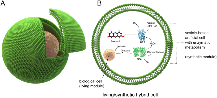
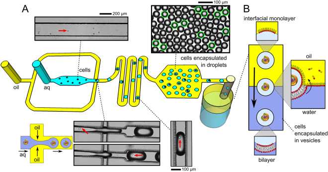
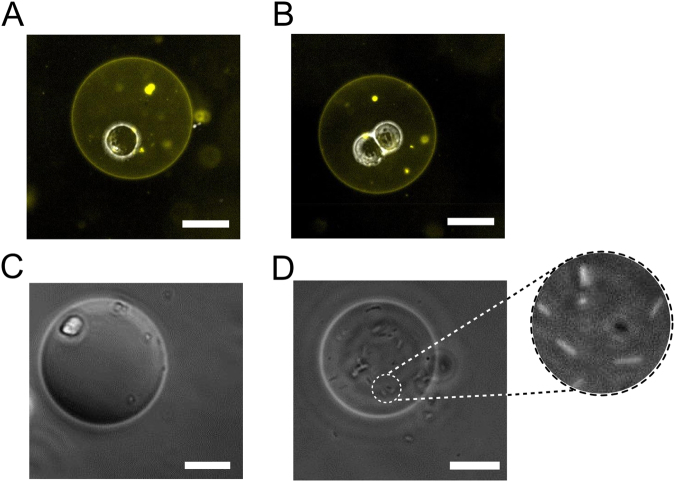
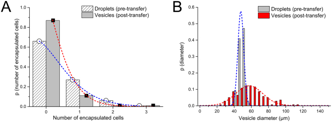
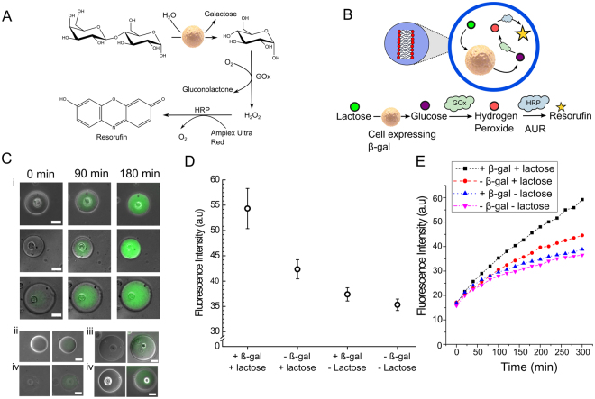
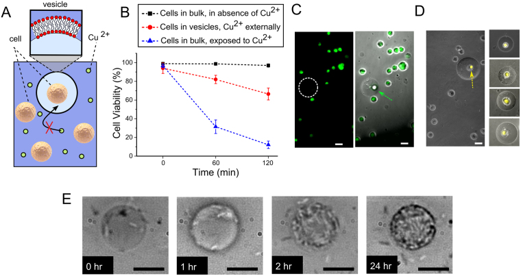
There is increasing interest in constructing artificial cells by functionalising lipid vesicles with biological and synthetic machinery. Due to their reduced complexity and lack of evolved biochemical pathways, the capabilities of artificial cells are limited in comparison to their biological counterparts. We show that encapsulating living cells in vesicles provides a means for artificial cells to leverage cellular biochemistry, with the encapsulated cells serving organelle-like functions as living modules inside a larger synthetic cell assembly. Using microfluidic technologies to construct such hybrid cellular bionic systems, we demonstrate that the vesicle host and the encapsulated cell operate in concert. The external architecture of the vesicle shields the cell from toxic surroundings, while the cell acts as a bioreactor module that processes encapsulated feedstock which is further processed by a synthetic enzymatic metabolism co-encapsulated in the vesicle.
Conflict of interest statement
The authors declare no competing interests.
Figures
Similar articles
-
Recent Progress in Micro/Nanoreactors toward the Creation of Artificial Organelles.Adv Healthc Mater. 2018 Mar;7(5). doi: 10.1002/adhm.201700917. Epub 2017 Dec 4. Adv Healthc Mater. 2018. PMID: 29205928 Review.
-
Bottom-Up Assembly of Functional Intracellular Synthetic Organelles by Droplet-Based Microfluidics.Small. 2020 Jul;16(27):e1906424. doi: 10.1002/smll.201906424. Epub 2020 Feb 20. Small. 2020. PMID: 32078238
-
Vesicle-based artificial cells as chemical microreactors with spatially segregated reaction pathways.Nat Commun. 2014 Oct 29;5:5305. doi: 10.1038/ncomms6305. Nat Commun. 2014. PMID: 25351716
-
Multivesicular droplets: a cell model system to study compartmentalised biochemical reactions.Lab Chip. 2017 Sep 12;17(18):3112-3119. doi: 10.1039/c7lc00710h. Lab Chip. 2017. PMID: 28813055 Free PMC article.
-
Microfluidic Handling and Analysis of Giant Vesicles for Use as Artificial Cells: A Review.Adv Biosyst. 2019 Jun;3(6):e1800318. doi: 10.1002/adbi.201800318. Epub 2019 May 7. Adv Biosyst. 2019. PMID: 32648705 Review.
Cited by
-
DNA-Based Optical Quantification of Ion Transport across Giant Vesicles.ACS Nano. 2022 Oct 25;16(10):17128-17138. doi: 10.1021/acsnano.2c07496. Epub 2022 Oct 12. ACS Nano. 2022. PMID: 36222833 Free PMC article.
-
Interfacing Living and Synthetic Cells as an Emerging Frontier in Synthetic Biology.Angew Chem Int Ed Engl. 2021 Mar 8;60(11):5602-5611. doi: 10.1002/anie.202006941. Epub 2020 Oct 13. Angew Chem Int Ed Engl. 2021. PMID: 32909663 Free PMC article. Review.
-
Formation of Polarized, Functional Artificial Cells from Compartmentalized Droplet Networks and Nanomaterials, Using One-Step, Dual-Material 3D-Printed Microfluidics.Adv Sci (Weinh). 2019 Oct 24;7(1):1901719. doi: 10.1002/advs.201901719. eCollection 2020 Jan. Adv Sci (Weinh). 2019. PMID: 31921557 Free PMC article.
-
Evolution and synthetic biology.Curr Opin Microbiol. 2023 Dec;76:102394. doi: 10.1016/j.mib.2023.102394. Epub 2023 Oct 4. Curr Opin Microbiol. 2023. PMID: 37801925 Free PMC article. Review.
-
Sculpting and fusing biomimetic vesicle networks using optical tweezers.Nat Commun. 2018 May 14;9(1):1882. doi: 10.1038/s41467-018-04282-w. Nat Commun. 2018. PMID: 29760422 Free PMC article.
References
-
- Stano P, Carrara P, Kuruma Y, de Souza TP, Luisi PL. Compartmentalized reactions as a case of soft-matter biotechnology: synthesis of proteins and nucleic acids inside lipid vesicles. Journal of Materials Chemistry. 2011;21:18887–18902. doi: 10.1039/c1jm12298c. - DOI
Publication types
MeSH terms
Substances
LinkOut - more resources
Full Text Sources
Other Literature Sources
