. 2018 Mar 14;9(3):404.
doi: 10.1038/s41419-018-0437-9.

Kir6.1/K-ATP channel modulates microglia phenotypes: implication in Parkinson's disease

Affiliations

Kir6.1/K-ATP channel modulates microglia phenotypes: implication in Parkinson's disease

Ren-Hong Du et al. Cell Death Dis. .

Abstract

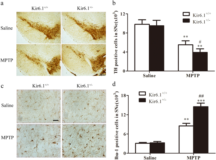
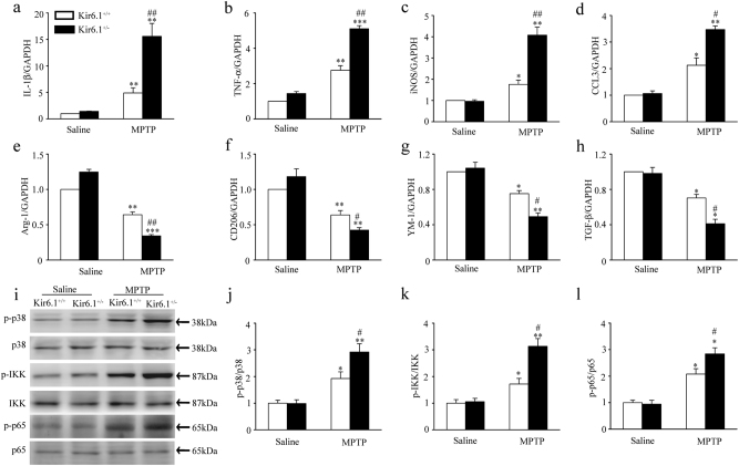
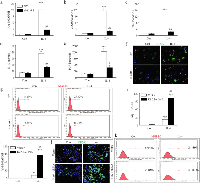
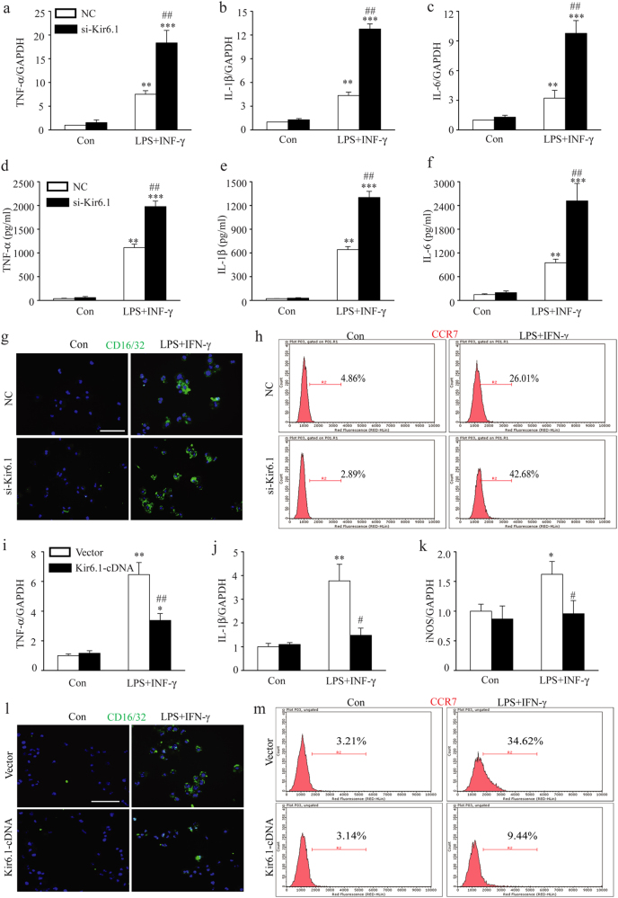
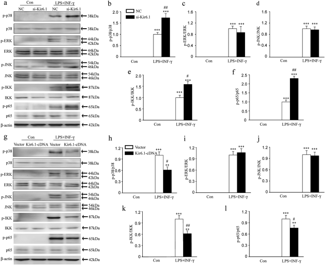
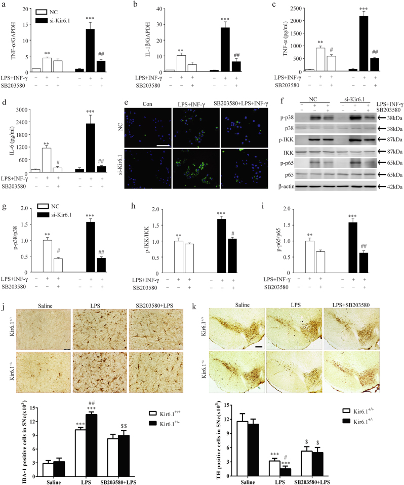
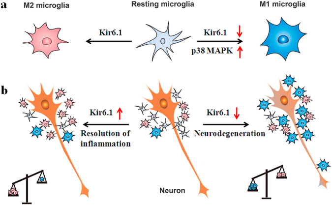
Classical activation (M1 phenotype) and alternative activation (M2 phenotype) are the two polars of microglial activation states that can produce either neurotoxic or neuroprotective effects in the immune pathogenesis of Parkinson's disease (PD). Exploiting the beneficial properties of microglia cells by modulating their polarization states provides great potential for the treatment of PD. However, the mechanism that regulates microglia polarization remains elusive. Here we demonstrated that Kir6.1-containing ATP-sensitive potassium (Kir6.1/K-ATP) channel switched microglia from the detrimental M1 phenotype toward the beneficial M2 phenotype. Kir6.1 knockdown inhibited M2 polarization and simultaneously exaggerated M1 microglial inflammatory responses, while Kir6.1 overexpression promoted M2 polarization and synchronously alleviated the toxic phase of M1 microglia polarization. Furthermore, we observed that the Kir6.1 deficiency dramatically exacerbated dopaminergic neuron death companied by microglia activation in mouse model of PD. Mechanistically, Kir6.1 deficiency enhanced the activation of p38 MAPK-NF-κB pathway and increased the ratio of M1/M2 markers in the substantia nigra compacta of mouse model of PD. Suppression of p38 MAPK in vivo partially rescued the deleterious effects of Kir6.1 ablation on microglia phenotype and dopaminergic neuron death. Collectively, our findings reveal that Kir6.1/K-ATP channel modulates microglia phenotypes transition via inhibition of p38 MAPK-NF-κB signaling pathway and Kir6.1/K-ATP channel may be a promising therapeutic target for PD.


Conflict of interest statement

The authors declare that they have no conflict of interest.

Figures

Fig. 1
Fig. 1. Kir6.1 deletion aggravated dopaminergic neuron loss and microglia overactivation in MPTP Parkinson’s disease model mice.
a Microphotographs of Tyrosine hydroxylase (TH)-positive neurons in the substantia nigra compacta (SNc). b Stereological counts of TH-positive neurons in the SNc. c Microphotographs of ionized calcium-binding adaptor molecule 1 (IBA-1)-positive cells in the SNc. d Stereological counts of IBA-1-positive cells in the SNc. Data are presented as mean ± SEM, **p < 0.01, ***p < 0.001 versus corresponding control (saline) group; #p < 0.05, ##p < 0.01 versus MPTP-treated Kir6.1+/+ groups. n = 6 for each group. MPTP 1-methyl-4-phenyl-1, 2, 3, 6-tetrahydropyridine
Fig. 2
Fig. 2. Kir6.1 deficiency facilitated the switch of microglia phenotypes from M2 to M1 in MPTP Parkinson’s disease model mice.
ah Kir6.1 deficiency increased the production of IL-1β (a), TNF−α (b), iNOS (c), and CCL3 (d), and reduced the expression of Arginase1 (e), CD206 (f), YM-1 (g) and TGF-β (h) in the substantia nigra compacta (SNc). il Kir6.1 deficiency enhanced the activation of p38 and NF-κB in the SNc. Representative immunoblot (i) and quantitative analysis of the phosphorylation of p38 (j), IKK (k) and p65 (l) in the SNc of Kir6.1+/+ and Kir6.1+/ mice. Data are presented as mean ± SEM, *p < 0.05, **p < 0.01, ***p < 0.001 versus corresponding control (saline) group; #p < 0.05, ##p < 0.01 versus MPTP-treated Kir6.1+/+ groups. n = 5 for each group. MPTP 1-methyl-4-phenyl-1, 2, 3, 6-tetrahydropyridine
Fig. 3
Fig. 3. Kir6.1 promoted M2 microglia polarization.
ag Kir6.1 knockdown reduced the expression of related M2 markers. The expression of Arginase1 (a), CD206 (b), YM-1 (c), IL-10 (d) and TGF-β (e) were assessed by qPCR or ELISA in microglia. f Representative immunofluorescence staining of CD206 was visualized under microscopy. g The expression of M2 related biomarker MGL1/2 was detected by flow cytometry. Data are presented as mean ± SEM from four independent experiments, ***p < 0.001 versus corresponding control; #p < 0.05, ##p < 0.01 versus IL-4-treated negative control (NC) groups. hk Kir6.1 overexpression increased the expression of related M2 markers. The expression of Arginase1 (h) and YM-1 (i) were assessed by qPCR. j Representative immunofluorescence staining of CD206 was visualized under microscopy. k The expression of M2 related biomarker MGL1/2 was detected by flow cytometry. Data are presented as mean ± SEM from four independent experiments, **p < 0.01, ***p < 0.001 versus corresponding control; ##p < 0.01 versus IL-4-treated vector groups
Fig. 4
Fig. 4. Kir6.1 inhibited M1 microglia polarization.
ah Kir6.1 knockdown increased the expression of related M1 markers. The expression of TNF-α, IL-1β and IL-6 were assessed by qPCR (ac) or ELISA (df) in BV2 microglia. g Representative immunofluorescence staining of CD16/32 was visualized under microscopy. h The expression of M1 related biomarker CCR7 was detected by flow cytometry. Data are presented as mean ± SEM from four independent experiments, **p < 0.01, ***p < 0.001 versus corresponding control; ##p < 0.01 versus LPS+INF-γ-treated negative control (NC) groups. im Kir6.1 overexpression inhibited the expression of related M1 markers. The expression of TNF-α (i), IL-1β (j) and iNOS (k) were assessed by qPCR. l Representative immunofluorescence staining of CD16/32 was visualized under microscopy. m The expression of M1 related biomarker CCR7 was detected by flow cytometry. Data are presented as mean ± SEM from four independent experiments, *p < 0.05, **p < 0.01 versus corresponding control; #p < 0.05, ##p < 0.01 versus LPS+INF-γ-treated vector groups
Fig. 5
Fig. 5. Kir6.1 inhibited the activation of p38 MAPK and NF-κB in microglia.
af Kir6.1 knockdown enhanced the phosphorylation of p38, IKK and p65 in microglia treated with LPS+INF-γ. Representative Immunoblot (a) and quantitative analysis of the phosphorylation of p38 (b), ERK (c), JNK (d), IKK (e) and p65 (f) in microglia. Data are presented as mean ± SEM from four independent experiments, ***p < 0.001 versus corresponding control; #p < 0.05, ##p < 0.01 versus LPS+INF-γ-treated negative control (NC) groups. gl Kir6.1 overexpression inhibited the phosphorylation of p38, IKK and p65 in microglia treated with LPS+INF-γ. Representative immunoblot (g) and quantitative analysis of the phosphorylation of p38 (h), ERK (i), JNK (j), IKK (k) and p65 (l) in microglia. Data are presented as mean ± SEM from four independent experiments, **p < 0.01, ***p < 0.001 versus corresponding control; #p < 0.05, ##p < 0.01 versus LPS+INF-γ-treated vector groups
Fig. 6
Fig. 6. Suppression of p38 MAPK inhibited M1 microglia polarization and alleviated dopaminergic neuron degeneration in Kir6.1-deficent mice of Parkinson’s disease model.
ad SB203580 reduced the expression of TNF-α (a, c), IL-1β (b) and IL-6 (d) were assessed by qPCR or ELISA in Kir6.1- knockdown microglia. e SB203580 reduced the expression of CD16/32 in Kir6.1 knockdown microglia. fi SB203580 suppressed the phosphorylation of p38, IKK and p65 in Kir6.1 knockdown microglia. Representative immunoblot (f) and quantitative analysis of the phosphorylation of p38 (g), IKK (h) and p65 (i) in microglia. Data are presented as mean ± SEM from four independent experiments, **p < 0.01, ***p < 0.001 versus corresponding control; #p < 0.05, ##p < 0.01 versus corresponding LPS+INF-γ groups. j-k SB203580 attenuated microglia activation and dopaminergic neuron loss in Kir6.1+/− mice of lipopolysaccharide-induced Parkinson’s disease model. Microphotographs and stereological counts of IBA-1-positive cells (j) and TH-positive neurons (k) in the substantia nigra compacta. Data are presented as mean ± SEM, ***p < 0.001 versus corresponding control (saline) group; #p < 0.05, ##p < 0.01 versus LPS-treated Kir6.1+/+ groups; $p < 0.05, $$p < 0.01 versus LPS-treated Kir6.1+/- groups. n = 4 for each group
Fig. 7
Fig. 7. Proposed model of Kir6.1/K-ATP channel involved in regulation of microglia phenotypes and neurodegeneration.
a Under physiological conditions, microglia exist in a resting state with ramified morphology. They dramatically polarize into M1 or M2 phenotype upon different immunological stimuli or injury. Kir6.1/K-ATP channel switches microglia from the detrimental M1 phenotype toward the beneficial M2 phenotype. Suppression of Kir6.1/K-ATP channel inhibits M2 polarization and exaggerates M1 polarization via activation of p38 MAPK. b Suppression of Kir6.1/K-ATP channel compromises neuroprotective effects of M2 microglia and accordingly exaggerates detrimental effects of M1 microglia. The increased ratio of M1/M2 microglia contributes to extensive neuron death and aggravates neurodegeneration

Similar articles

Cited by

References

    1. Engelender S, Isacson O. The threshold theory for Parkinson’s disease. Trends Neurosci. 2017;40:4–14. doi: 10.1016/j.tins.2016.10.008. - DOI - PubMed
    1. Pal R, Tiwari PC, Nath R, Pant KK. Role of neuroinflammation and latent transcription factors in pathogenesis of Parkinson’s disease. Neurol. Res. 2016;38:1111–1122. doi: 10.1080/01616412.2016.1249997. - DOI - PubMed
    1. Zhou Y, et al. MicroRNA-7 targets Nod-like receptor protein 3 inflammasome to modulate neuroinflammation in the pathogenesis of Parkinson’s disease. Mol. Neurodegener. 2016;11:28. doi: 10.1186/s13024-016-0094-3. - DOI - PMC - PubMed
    1. Walker DG, Lue LF. Immune phenotypes of microglia in human neurodegenerative disease: challenges to detecting microglial polarization in human brains. Alzheimers Res. Ther. 2015;7:56. doi: 10.1186/s13195-015-0139-9. - DOI - PMC - PubMed
    1. Colton C, Wilcock DM. Assessing activation states in microglia. CNS Neurol. Disord. Drug Targets. 2010;9:174–191. doi: 10.2174/187152710791012053. - DOI - PubMed

Publication types