

Pseudo-hemorrhagic region formation in pancreatic neuroendocrine tumors is a result of blood vessel dilation followed by endothelial cell detachment
- PMID: 29541192
- PMCID: PMC5835859
- DOI: 10.3892/ol.2018.7840
Pseudo-hemorrhagic region formation in pancreatic neuroendocrine tumors is a result of blood vessel dilation followed by endothelial cell detachment
Abstract
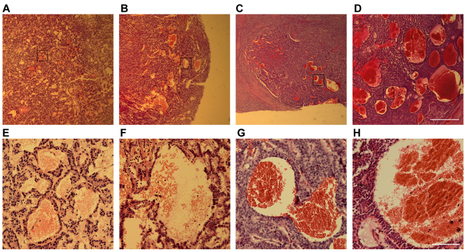
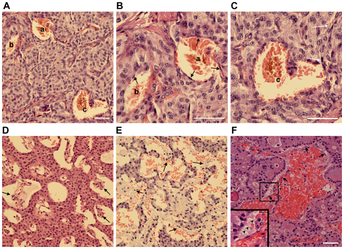
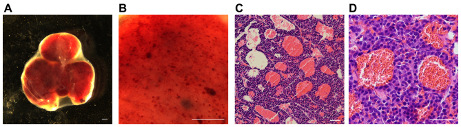
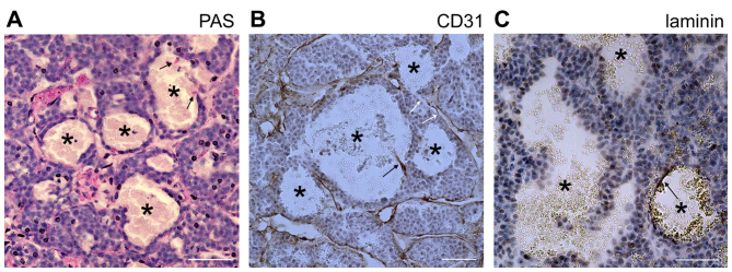
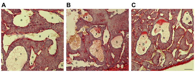
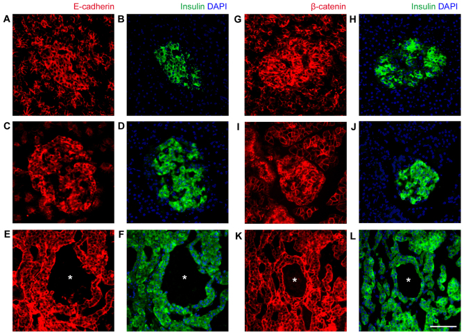
Aberrant blood vessel formation and hemorrhage may contribute to tumor progression and are potential targets in the treatment of several types of cancer. Pancreatic neuroendocrine tumors (PNETs) are highly vascularized, particularly when they are well-differentiated. However, the process of vascularization and endothelial cell detachment in PNETs is poorly understood. In the present study, 132 PNET clinical samples were examined and a special type of hemorrhagic region was observed in ~30% of the samples regardless of tumor subtype. These hemorrhagic regions were presented as blood-filled caverns with a smooth boundary and were unlined by endothelial cells. Based on the extensive endothelial cell detachment observed in the clinical samples, the formation process of these blood-filled caverns was hypothesized. Blood vessel dilation followed by detachment of endothelial cells from the surrounding tumor tissue was speculated. This was further supported using an INS-1 xenograft insulinoma model. As the formation process was distinct from the typical diffusive hemorrhage, it was named 'pseudo-hemorrhage'. Furthermore, it was demonstrated that epithelial (E-) cadherin and β-catenin were overexpressed in tumor cells surrounding these pseudo-hemorrhagic regions. Therefore, even though no statistically significant association of pseudo-hemorrhage with clinical features (metastasis or disease recurrence) was identified, the high levels of E-cadherin and β-catenin expression may suggest that a number of features of normal islet cells are retained.
Keywords: INS-1 cells; endothelial cells; epithelial cadherin; hemorrhage; pancreatic neuroendocrine tumor; β-catenin.
Figures
Similar articles
-
Impact of Snail and E-cadherin expression in pancreatic neuroendocrine tumors.Oncol Lett. 2017 Aug;14(2):1697-1702. doi: 10.3892/ol.2017.6306. Epub 2017 Jun 2. Oncol Lett. 2017. PMID: 28789397 Free PMC article.
-
Alterations of E-cadherin, alpha-catenin and beta-catenin expression in neuroendocrine tumors of the gastrointestinal tract.Virchows Arch. 2002 Feb;440(2):145-54. doi: 10.1007/s004280100529. Virchows Arch. 2002. PMID: 11964044
-
Alteration of the E-cadherin/beta-catenin cell adhesion system is common in pulmonary neuroendocrine tumors and is an independent predictor of lymph node metastasis in atypical carcinoids.Cancer. 2005 Mar 15;103(6):1154-64. doi: 10.1002/cncr.20901. Cancer. 2005. PMID: 15712207
-
The up-to-date review of epidemiological pancreatic neuroendocrine tumors in Japan.J Hepatobiliary Pancreat Sci. 2015 Aug;22(8):574-7. doi: 10.1002/jhbp.225. Epub 2015 Feb 16. J Hepatobiliary Pancreat Sci. 2015. PMID: 25689058 Review.
-
Antiangiogenic Therapy in Pancreatic Neuroendocrine Tumors.Anticancer Res. 2016 Oct;36(10):5025-5030. doi: 10.21873/anticanres.11071. Anticancer Res. 2016. PMID: 27798861 Review.
Cited by
-
Apatinib inhibits tumor growth and angiogenesis in PNET models.Endocr Connect. 2019 Jan 1;8(1):8-19. doi: 10.1530/EC-18-0397. Endocr Connect. 2019. PMID: 30557852 Free PMC article.
-
The Role of the Tumor Microenvironment in Gastroenteropancreatic Neuroendocrine Tumors.Int J Mol Sci. 2025 Jun 12;26(12):5635. doi: 10.3390/ijms26125635. Int J Mol Sci. 2025. PMID: 40565098 Free PMC article. Review.
-
The Landscape and Clinical Application of the Tumor Microenvironment in Gastroenteropancreatic Neuroendocrine Neoplasms.Cancers (Basel). 2022 Jun 13;14(12):2911. doi: 10.3390/cancers14122911. Cancers (Basel). 2022. PMID: 35740577 Free PMC article. Review.
References
-
- Daldrup H, Shames DM, Wendland M, Okuhata Y, Link TM, Rosenau W, Lu Y, Brasch RC. Correlation of dynamic contrast-enhanced MR imaging with histologic tumor grade: Comparison of macromolecular and small-molecular contrast media. AJR Am J Roentgenol. 1998;171:941–949. doi: 10.2214/ajr.171.4.9762973. - DOI - PubMed
LinkOut - more resources
Full Text Sources
Other Literature Sources
Research Materials