. 2018 Apr;15(4):4683-4689.
doi: 10.3892/ol.2018.7848. Epub 2018 Jan 24.

Microrna-136 promotes proliferation and invasion ingastric cancer cells through Pten/Akt/P-Akt signaling pathway

Affiliations

Microrna-136 promotes proliferation and invasion ingastric cancer cells through Pten/Akt/P-Akt signaling pathway

Xuyan Chen et al. Oncol Lett. 2018 Apr.

Abstract

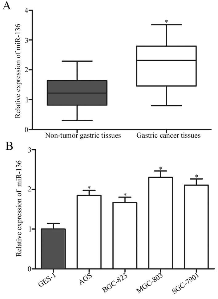
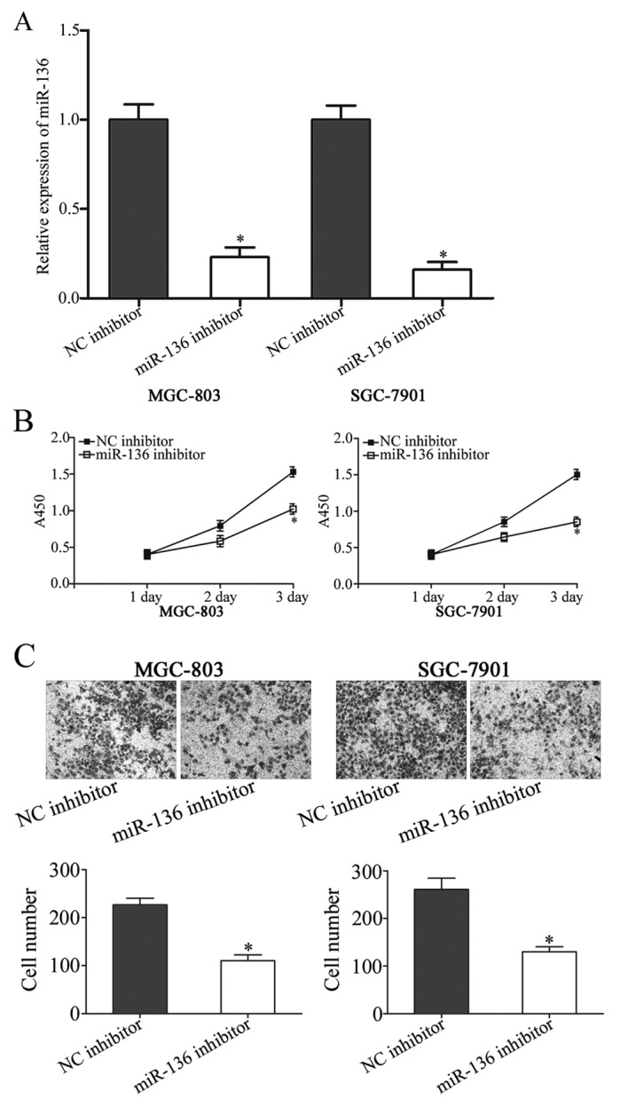
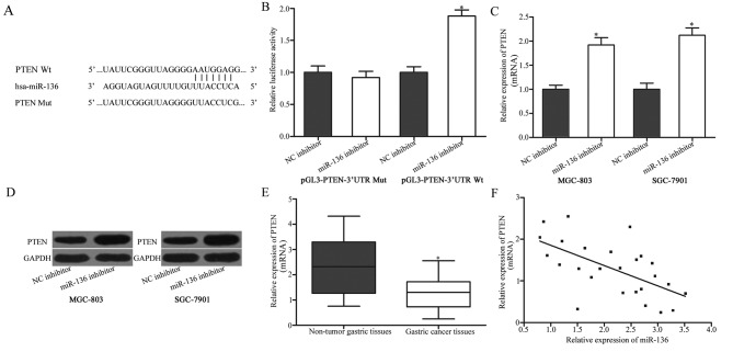
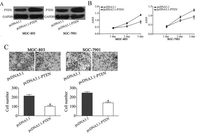
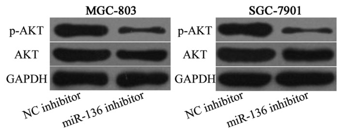
Gastric cancer is the fourth most common cancer and the second most frequent cause of cancer-associated mortality in the world. Previous studies have revealed that expression levels of microRNAs (miRNAs) are associated with the initiation and progression of several types of cancer, including gastric cancer. Previous studies have demonstrated that the abnormal expression of miRNA-136 may serve a function in the progression of several types of human cancer. However, the expression pattern of miR-136, its functions and underlying molecular mechanisms in gastric cancer remain unresolved. In the present study, it was revealed that the expression of miR-136 was aberrantly up regulated in gastric cancer tissues and cell lines. The suppression of miR-136 was able to inhibit proliferation and invasion in gastric cancer cell lines. Furthermore, phosphatase and tensin homolog (PTEN) was identified as a direct target gene of miR-136 in gastric cancer. PTEN was under expressed in gastric cancer tissues compared with non-tumor gastric tissues, and PTEN expression was negatively correlated with miR-136 expression. Furthermore, PTEN overexpression mimics the effects of miR-136 knockdown on gastric cancer cells. Additionally, miR-136 under expression decreased phospho-(p) AKT expression, but did not affect AKT expression in gastric cancer cells. In conclusion, the data of the present study suggest that miR-136 acts as an oncogene in gastric cancer via regulation of the PTEN/AKT/p-AKT signaling pathway and may potentially serve as a novel therapeutic target for the treatment of gastric cancer.

Keywords: gastric cancer; invasion; microRNA-136; phosphatase and tensin homolog; proliferation.


Figures

Figure 1.
Figure 1.
Reverse transcription-quantitative polymerase chain reaction analysis of miR-136 expression in gastric cancer tissues and cell lines. (A) miR-136 expression was determined in 26 paired gastric cancer tissues and matched adjacent non-tumorous gastric mucosae tissues. The data were presented as box plots. The top of the box indicates upper quartile, and the bottom of the box indicates lower quartile. The central line in the box indicates median, and the whiskers indicate the range. *P<0.05 vs. non-tumor tissues (B) Relative expression of miR-136 in four gastric cancer cell lines (AGS, BGC-823, MGC-803 and SGC-7901) and normal human gastric epithelial GES-1 cells. Data are presented as the mean ± standard deviation. *P<0.05 vs. GES-1. miR, microRNA.
Figure 2.
Figure 2.
miR-136 knockdown inhibits proliferation and invasion of gastric cancer cells. (A) The expression level of miR-136 in MGC-803 and SGC-7901 cells that were treated with miR-136 inhibitor or NC inhibitor. (B) A cell proliferation assay was performed at 1, 2 and 3 days following the plating of transfected cells. (C) A Transwell cell invasion assay of MGC-803 and SGC-7901 cells that were transfected with miR-136 inhibitor or NC inhibitor. Magnification, ×200. Data are presented as the mean ± standard deviation. *P<0.05 vs. NC inhibitor. miR, microRNA; NC, negative control.
Figure 3.
Figure 3.
PTEN is a direct target of miR-136. (A) Binding sequences for miR-136 in the 3′UTR of PTEN, and the mutations in the 3′UTR of PTEN are presented. (B) A luciferase reporter assay was performed in 293T cells at 48 h after co-transfection with miR-136 inhibitor or NC inhibitor, and pGL3-PTEN-3′UTR Wt or pGL3-PTEN-3′UTR Mut. The treatment with a miR-136 inhibitor was able to increase luciferase activity in cells that were transfected with pGL3-PTEN-3′UTR Wt. By contrast, miR-136 inhibitor did not affect the luciferase activity in in cells that were transfected with pGL3-PTEN-3′UTR Mut. *P<0.05 vs. NC inhibitor. (C) RT-qPCR analysis of PTEN mRNA in MGC-803 and SGC-7901 cells following transfection with miR-136 inhibitor or NC inhibitor. *P<0.05 vs. NC inhibitor. (D) Western blotting was used to detect PTEN protein expression in MGC-803 and SGC-7901 cells following transfection with miR-136 inhibitor or NC inhibitor. (E) The relative PTEN mRNA expression levels were determined using RT-qPCR in gastric cancer tissues and adjacent non-tumorous gastric mucosae tissues. The data are presented as box plots. The top of the box indicates the upper quartile, and the bottom indicates the lower quartile. The central line in the box indicates median, and the whiskers indicate the range. *P<0.05 vs. non-tumorous gastric mucosae tissues. (F) Analysis of correlation between miR-136 and PTEN mRNA expression in gastric cancer tissues. r=−0.6035, P=0.0011. The data are presented as the mean ± standard deviation. PTEN, phosphatase and tensin homolog deleted on chromosome ten; miR-136, microRNA-136; NC inhibitor, negative control inhibitor; 3′UTR, 3′-untranslated regions; Wt, wild-type; Mut, mutant; RT-qPCR, reverse transcription-quantitative polymerase chain reaction.
Figure 4.
Figure 4.
Up regulation of PTEN mimics the effects of miR-136 under expression in gastric cancer cell proliferation and invasion. (A) Western blot analysis of PTEN expression in MGC-803 and SGC-7901 cells that were treated with pcDNA3.1-PTEN or pcDNA3.1. (B) Cell proliferation assay analysis of cell proliferation following pcDNA3.1-PTEN or pcDNA3.1 transfection in MGC-803 and SGC-7901 cells. (C) Transwell cell invasion assay was performed to detect the effects on cell invasion following pcDNA3.1-PTEN or pcDNA3.1 transfection in MGC-803 and SGC-7901 cells. The data are presented as the mean ± standard deviation. *P<0.05 vs. pcDNA3.1. PTEN, phosphatase and tensin homolog.
Figure 5.
Figure 5.
miR-136 knockdown decreases p-AKT expression, but not AKT expression in MGC-803 and SGC-7901 cells. AKT, protein kinase B. p-, phosphorylated; miR-136, microRNA-136; NC, negative control.

References

    1. Jemal A, Bray F, Center MM, Ferlay J, Ward E, Forman D. Global cancer statistics. CA Cancer J Clin. 2011;61:69–90. doi: 10.3322/caac.20107. - DOI - PubMed
    1. Yang L. Incidence and mortality of gastric cancer in China. World J Gastroenterol. 2006;12:17–20. doi: 10.3748/wjg.v12.i1.17. - DOI - PMC - PubMed
    1. Guo M, Yan W. Epigenetics of gastric cancer. Methods Mol Biol. 2015;1238:783–799. doi: 10.1007/978-1-4939-1804-1_41. - DOI - PubMed
    1. Ueda T, Volinia S, Okumura H, Shimizu M, Taccioli C, Rossi S, Alder H, Liu CG, Oue N, Yasui W, et al. Relation between microRNA expression and progression and prognosis of gastric cancer: A microRNA expression analysis. Lancet Oncol. 2010;11:136–146. doi: 10.1016/S1470-2045(09)70343-2. - DOI - PMC - PubMed
    1. Terry MB, Gaudet MM, Gammon MD. The epidemiology of gastric cancer. Semin Radiat Oncol. 2002;12:111–127. doi: 10.1053/srao.30814. - DOI - PubMed