. 2017 Nov 6;9(13):10891-10904.
doi: 10.18632/oncotarget.22443. eCollection 2018 Feb 16.

SOX5 predicts poor prognosis in lung adenocarcinoma and promotes tumor metastasis through epithelial-mesenchymal transition

Affiliations

SOX5 predicts poor prognosis in lung adenocarcinoma and promotes tumor metastasis through epithelial-mesenchymal transition

Xin Chen et al. Oncotarget. .

Abstract

Lung cancer is the leading cause of cancer-related death worldwide. Epithelial-mesenchymal transition (EMT) promotes lung cancer progression and metastasis, especially in lung adenocarcinoma. Sex determining region Y-box protein 5 (SOX5) is known to stimulate the progression of various cancers. Here, we used immunohistochemical analysis to reveal that SOX5 levels were increased in 90 lung adenocarcinoma patients. The high SOX5 expression in lung adenocarcinoma and non-tumor counterparts correlated with the patients' poor prognosis. Inhibiting SOX5 expression attenuated metastasis and progression in lung cancer cells, while over-expressing SOX5 accelerated lung adenocarcinoma progression and metastasis via EMT. An in vivo zebrafish xenograft cancer model also showed SOX5 knockdown was followed by reduced lung cancer cell proliferation and metastasis. Our results indicate SOX5 promotes lung adenocarcinoma tumorigenicity and can be a novel diagnosis and prognosis marker of the disease.

Keywords: EMT; SOX5; lung adenocarcinoma; prognosis.


Conflict of interest statement

CONFLICTS OF INTEREST The authors declare that they have no conflicts of interest.

Figures

Figure 1
Figure 1. SOX5 is over-expressed in lung adenocarcinoma and is associated with in vitro cell invasion
(A) Western blot analysis of SOX5 levels (top) in four human lung adenocarcinoma patients (LAC) and their respective non-tumor counterparts (N); Quantitative RT-PCR analysis of SOX5mRNA level (bottom) in the same patients, normalized versus GAPDH; ***:p < 0.001 (LAC vs. N). (B) Immunohistochemical analysis of SOX5expression and localization in LACs and paracancerous tissues (Microscope magnification: 200×). SOX5 was localized in the cytoplasm. (C) Western blot analysis of SOX5 level (top) in several lung cancer cell lines and bronchial epithelium cell line (16HBE); Quantitative RT-PCR analysis of SOX5 level in these cell lines (middle); Invasive capacity analysis in different cell lines (bottom), tested in the 8μm invasive chamber. Data represent mean ± SD calculated from triplicate experiments.
Figure 2
Figure 2. SOX5 (in both tumor and paracancerous tissues) correlates negatively with survival in LAC patients
(A) Overall survival rate presented in Kaplan−Meier survival curve for cases with high SOX5 expression versus cases with low SOX5 expression in 90 LAC patients’ cancerous tissues. There was significant difference in prognosis between these two groups (P < 0.05) (B) Kaplan−Meier survival curve analysis of SOX5 expression in the 90 LAC patients’ paracancerous tissues (P < 0.05).
Figure 3
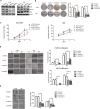
Figure 3. SOX5 knockdown inhibits LAC cell proliferation and metastasis in vitro
(A) SOX5 expression in control (Untreated), sh Negative Control (scramble sequence, NC), and SOX5 knockdown (shSOX5-1and shSOX5-2) LAC cells H1299 and 95D was detected by western blot. Cell cycle biomarkers CyclinD1 and c-Myc were also detected. (B) Colony formation analysis of shSOX5-1 and shSOX5-2, as well as untreated and negative control cells. Cells with stable shRNA expression were seeded into 6-well plates at 2000 cells per well. Seven days later, crystal violet staining and colony counting was performed. Data were shown on the right as mean ± SD and *** for P < 0.001 (LV-shNC vs. LV-shSOX5). (C) Effect of SOX5 knockdown on cell proliferation was determined by CCK8 assay at the indicated time point. ** for P < 0.01(LV-shNC vs. LV-shSOX5). (D) Effect of SOX5 knockdown on cell migration by wound-healing assay. Statistical analysis was done with GraphPad Prism 5, ** for P < 0.01 and *** for P < 0.001 (LV-shNC vs. LV-shSOX5). (E) Invasive transwell analysis of shSOX5-1 and shSOX5-2, as well as untreated and shNC cells. *** for P < 0.001 (LV-shNC vs. LV-shSOX5).
Figure 4
Figure 4. Effects of SOX5 overexpression on LAC cell proliferation and metastasis
(A) SOX5 expression in control (Vector) and SOX5 overexpression (SOX5) in A549 and H1975 cells were detected by western blot. CyclinD1 and c-Myc were also detected. (B) Effect of SOX5 overexpression on colony formation. A549 and H1975 cells with stable expression of vector or SOX5 were seeded into 6-well plates at 2000 cells per well and cultured for 7 days, followed by crystal violet staining and colony counting. Data were analyzed as mean ± SD and ** for P < 0.01(Right, Vector vs. SOX5). (C) Effect of SOX5 overexpression on the cell proliferation rate in A549 and H1975 cells expressing empty vector or SOX5 and determined by CCK8 assay at the indicated time point, * for P < 0.05 and ** for P < 0.01(Vector vs. SOX5). (D) Effect of SOX5 overexpression on cell migration in the wound-healing assay. Statistical analysis was done with GraphPad Prism 5, * for P < 0.05 and ** for P < 0.01(Vector vs. SOX5). (E) Invasive transwell analysis of SOX5 overexpression in A549 and H1975 cells. ** for P < 0.01(Vector vs. SOX5).
Figure 5
Figure 5. SOX5 facilitates the epithelial-mesenchymal transition (EMT) in lung adenocarcinoma
(A) Western blot analysis of epithelial (E-cadherin) and mesenchymal (N-cadherin , ZEB1, Vimentin) markers in control (Untreated), sh Negative Control (scramble sequence, NC) and SOX5 knockdown (shSOX5-1and shSOX5-2) LAC cells H1299 and 95D. (B) Western blot analysis of epithelial (E-cadherin) and mesenchymal (Vimentin and Twist1) markers in A549 and H1975 cells (lentivirus with empty vector or SOX5 transfected). (C) Immunohistochemical analysis of SOX5, E-cadherin, and Vimentin expression in three advanced-stage LAC patients with serial sections.
Figure 6
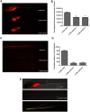
Figure 6. Silencing SOX5 reducesH1299 cell proliferation and metastasis in vivo
(A) Zebrafish cancer xenograft assay of H1299 cells with silenced SOX5. Arrows indicate metastatic tumor cells. (B) The integrated fluorescence intensity of tumors in the H1299-shSOX5 group compared with the H1299-NC group,**p < 0.01,***p < 0.001. (C) The metastasis inhibition of SOX5 knockdown. Arrows indicate metastatic tumor cells. (D) The cumulative distance of cell migration in the H1299-shSOX5 group compared with the H1299-NC group, **p < 0.01. (E) The schematic diagram of data collection and analysis in the zebrafish cancer xenograft model.

References

    1. Siegel RL, Miller KD, Jemal A. Cancer statistics, 2015. CA Cancer J Clin. 2015;65:5–29. https://doi.org/10.3322/caac.21254. - DOI - PubMed
    1. Chen W, Zheng R, Baade PD, Zhang S, Zeng H, Bray F, Jemal A, Yu XQ, He J. Cancer statistics in China, 2015. CA Cancer J Clin. 2016;66:115–32. https://doi.org/10.3322/caac.21338. - DOI - PubMed
    1. Miller KD, Siegel RL, Lin CC, Mariotto AB, Kramer JL, Rowland JH, Stein KD, Alteri R, Jemal A. Cancer treatment and survivorship statistics, 2016. CA Cancer J Clin. 2016;66:271–89. https://doi.org/10.3322/caac.21349. - DOI - PubMed
    1. Chen K, Zhou F, Shen W, Jiang T, Wu X, Tong X, Shao YW, Qin S, Zhou C. Novel mutations on EGFR Leu792 potentially correlate to acquired resistance to osimertinib in advanced NSCLC. J Thorac Oncol. 2017 https://doi.org/10.1016/j.jtho.2016.12.024. - DOI - PubMed
    1. Chambers AF, Groom AC, MacDonald IC. Dissemination and growth of cancer cells in metastatic sites. Nat Rev Cancer. 2002;2:563–72. https://doi.org/10.1038/nrc865. - DOI - PubMed

LinkOut - more resources