

Diffuse reflectance spectroscopy of human liver tumor specimens - towards a tissue differentiating optical biopsy needle using light emitting diodes
- PMID: 29541504
- PMCID: PMC5846514
- DOI: 10.1364/BOE.9.001069
Diffuse reflectance spectroscopy of human liver tumor specimens - towards a tissue differentiating optical biopsy needle using light emitting diodes
Abstract
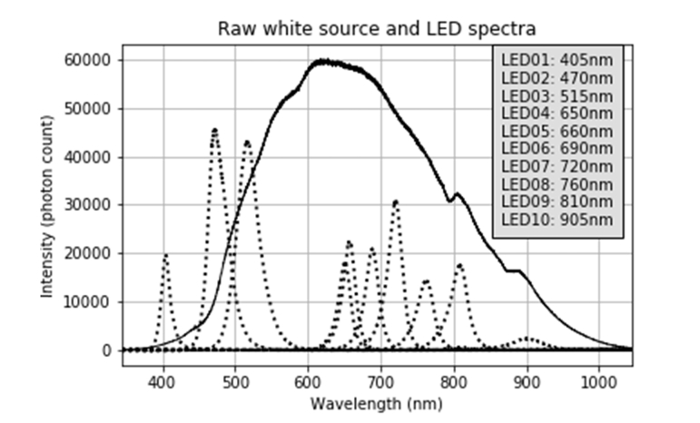
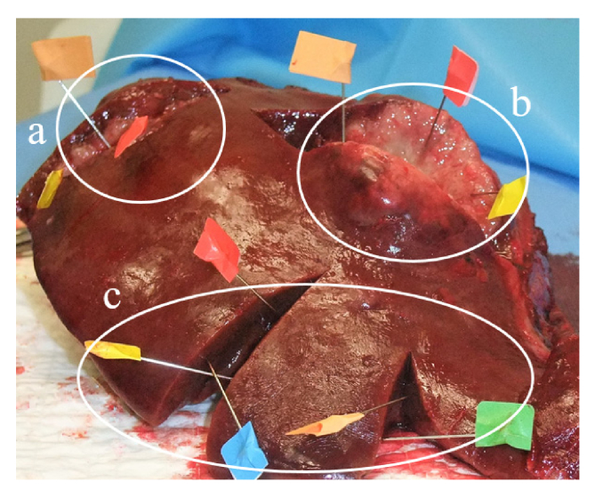
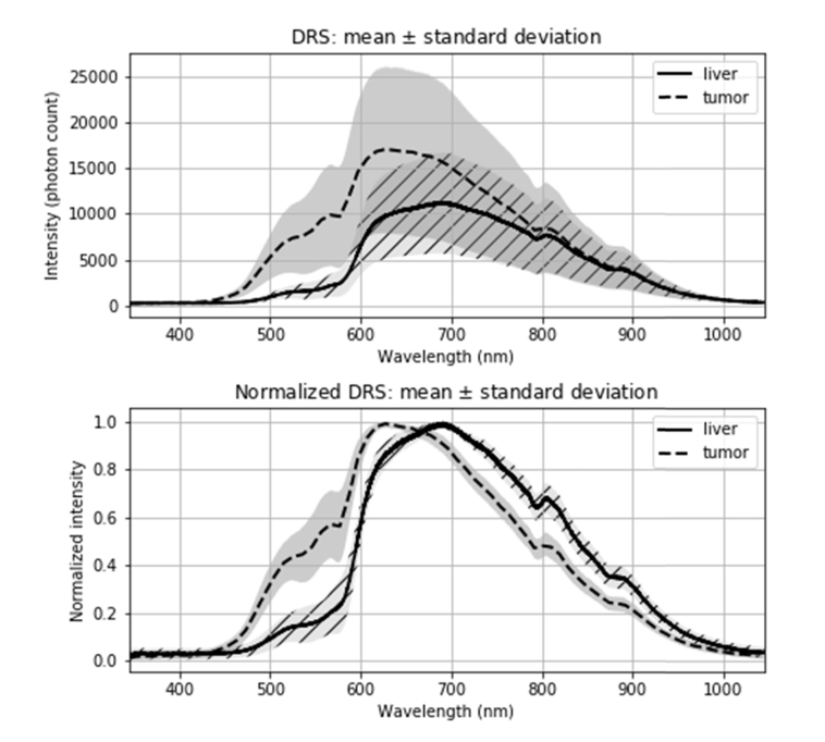
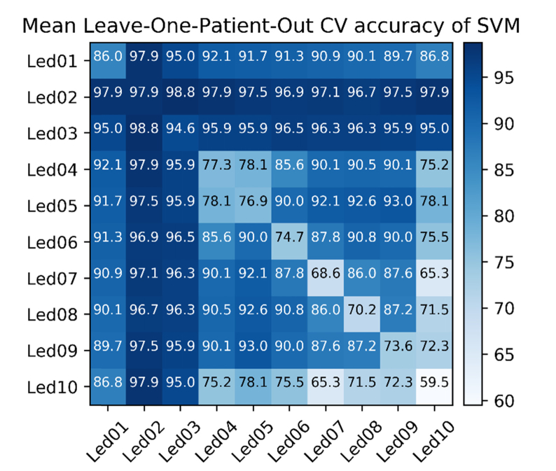
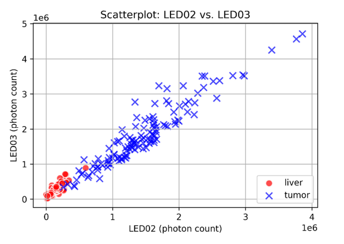
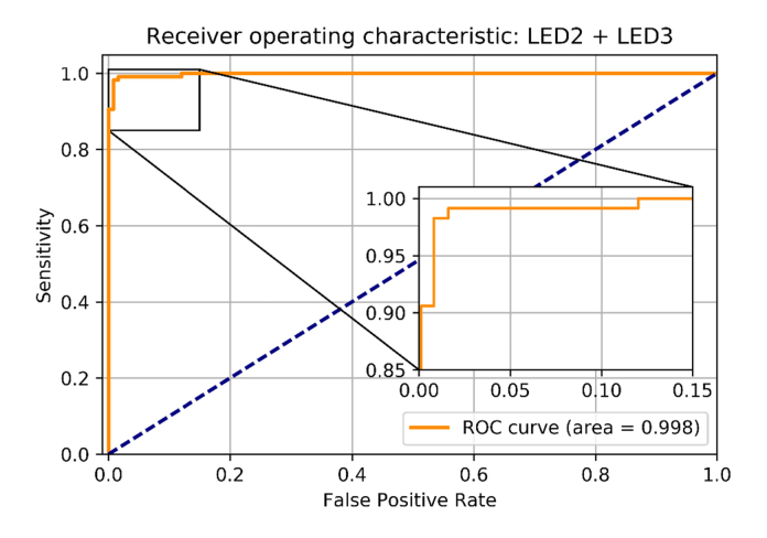
Significant numbers of liver biopsies fail to yield representative tissue samples. This study was conducted to evaluate the ability of LED-based diffuse reflectance spectroscopy to discriminate tumors from liver parenchyma. Ex vivo spectra were acquired from malignant lesions and liver parenchyma of 32 patients who underwent liver resection using a white light source and several LEDs. Integrated spectra of two combined LEDs with emission peaks at 470 nm and 515 nm were classified with 98.4% sensitivity and 99.2% specificity. The promising results could yield to a simple handheld and cost-efficient tool for real-time tissue differentiation implemented in a biopsy needle.
Keywords: (060.2310) Fiber optics; (170.1610) Clinical applications; (170.4580) Optical diagnostics for medicine; (170.6510) Spectroscopy, tissue diagnostics; (170.6935) Tissue characterization; (230.3670) Light-emitting diodes.
Conflict of interest statement
The authors declare that there are no conflicts of interest related to this article.
Figures
Similar articles
-
Human feasibility study of fluorescence spectroscopy guided optical biopsy needle for prostate cancer diagnosis.Annu Int Conf IEEE Eng Med Biol Soc. 2015;2015:7358-61. doi: 10.1109/EMBC.2015.7320091. Annu Int Conf IEEE Eng Med Biol Soc. 2015. PMID: 26737991
-
Towards the use of diffuse reflectance spectroscopy for real-time in vivo detection of breast cancer during surgery.J Transl Med. 2018 Dec 19;16(1):367. doi: 10.1186/s12967-018-1747-5. J Transl Med. 2018. PMID: 30567584 Free PMC article.
-
Optical spectroscopy characteristics can differentiate benign and malignant renal tissues: a potentially useful modality.J Urol. 2005 Nov;174(5):1754-8. doi: 10.1097/01.ju.0000177484.33596.c9. J Urol. 2005. PMID: 16217277
-
Dual-modality optical biopsy of glioblastomas multiforme with diffuse reflectance and fluorescence: ex vivo retrieval of optical properties.J Biomed Opt. 2017 Feb 1;22(2):27002. doi: 10.1117/1.JBO.22.2.027002. J Biomed Opt. 2017. PMID: 28157245
-
Role of In Vivo Reflectance Confocal Microscopy in the Analysis of Melanocytic Lesions.Acta Dermatovenerol Croat. 2018 Apr;26(1):64-67. Acta Dermatovenerol Croat. 2018. PMID: 29782304 Review.
Cited by
-
Model-based characterization platform of fiber optic extended-wavelength diffuse reflectance spectroscopy for identification of neurovascular bundles.J Biomed Opt. 2022 Sep;27(9):095002. doi: 10.1117/1.JBO.27.9.095002. J Biomed Opt. 2022. PMID: 36088529 Free PMC article.
-
Optical percutaneous needle biopsy of the liver: a pilot animal and clinical study.Sci Rep. 2020 Aug 26;10(1):14200. doi: 10.1038/s41598-020-71089-5. Sci Rep. 2020. PMID: 32848190 Free PMC article.
-
Real-time tracking of a diffuse reflectance spectroscopy probe used to aid histological validation of margin assessment in upper gastrointestinal cancer resection surgery.J Biomed Opt. 2022 Feb;27(2):025001. doi: 10.1117/1.JBO.27.2.025001. J Biomed Opt. 2022. PMID: 35106980 Free PMC article.
-
Optical characterization of the liver tissue affected by fibrolamellar hepatocellular carcinoma based on internal filters of laser-induced fluorescence.Sci Rep. 2022 Apr 12;12(1):6116. doi: 10.1038/s41598-022-10146-7. Sci Rep. 2022. PMID: 35414645 Free PMC article.
-
Point Projection Mapping System for Tracking, Registering, Labeling, and Validating Optical Tissue Measurements.J Imaging. 2024 Jan 30;10(2):37. doi: 10.3390/jimaging10020037. J Imaging. 2024. PMID: 38392085 Free PMC article.
References
-
- Francque S. M., De Pauw F. F., Van den Steen G. H., Van Marck E. A., Pelckmans P. A., Michielsen P. P., “Biopsy of focal liver lesions: guidelines, comparison of techniques and cost-analysis,” Acta Gastroenterol. Belg. 66(2), 160–165 (2003). - PubMed
-
- de Boer L. L., Hendriks B. H. W., van Duijnhoven F., Peeters-Baas M. T., Van de Vijver K., Loo C. E., Jóźwiak K., Sterenborg H. J. C. M., Ruers T. J. M., “Using DRS during breast conserving surgery: identifying robust optical parameters and influence of inter-patient variation,” Biomed. Opt. Express 7(12), 5188–5200 (2016).10.1364/BOE.7.005188 - DOI - PMC - PubMed
-
- Evers D. J., Nachabé R., Vranken Peeters M. J., van der Hage J. A., Oldenburg H. S., Rutgers E. J., Lucassen G. W., Hendriks B. H., Wesseling J., Ruers T. J., “Diffuse reflectance spectroscopy: towards clinical application in breast cancer,” Breast Cancer Res. Treat. 137(1), 155–165 (2013).10.1007/s10549-012-2350-8 - DOI - PubMed
-
- Nachabé R., Evers D. J., Hendriks B. H., Lucassen G. W., van der Voort M., Rutgers E. J., Peeters M. J., Van der Hage J. A., Oldenburg H. S., Wesseling J., Ruers T. J., “Diagnosis of breast cancer using diffuse optical spectroscopy from 500 to 1600 nm: comparison of classification methods,” J. Biomed. Opt. 16(8), 087010 (2011).10.1117/1.3611010 - DOI - PubMed
LinkOut - more resources
Full Text Sources
Other Literature Sources