

Two Cases of Myiasis Associated with Malignancies in Patients Living in the Continental United States
- PMID: 29541570
- PMCID: PMC5846802
- DOI: 10.7759/cureus.2049
Two Cases of Myiasis Associated with Malignancies in Patients Living in the Continental United States
Abstract
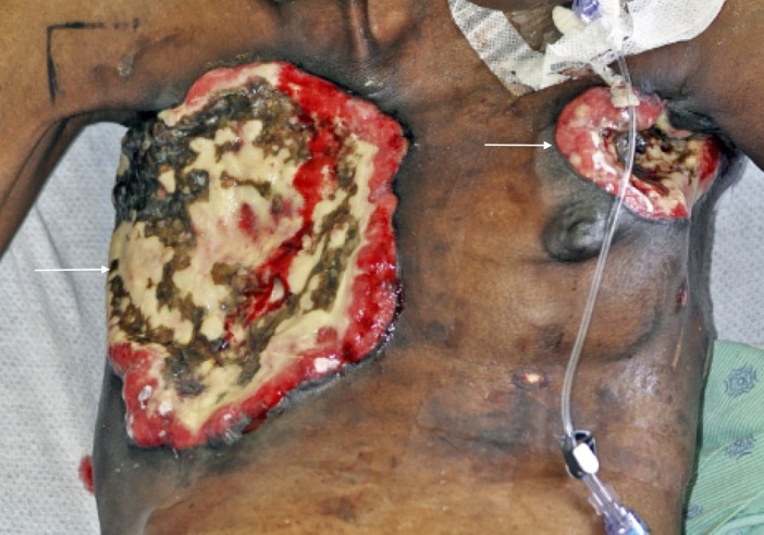
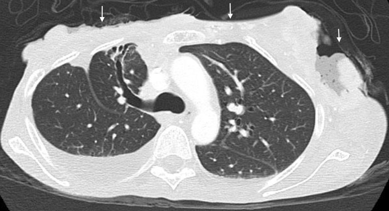
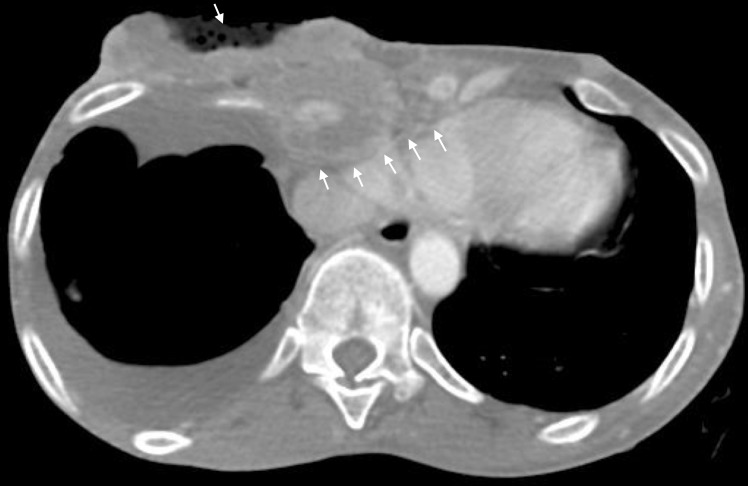
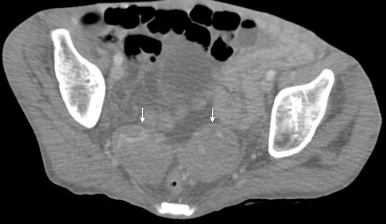
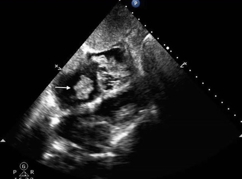
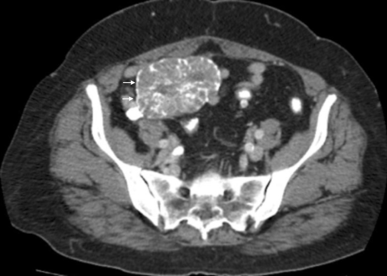
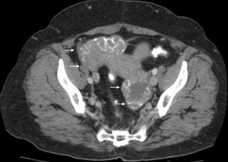
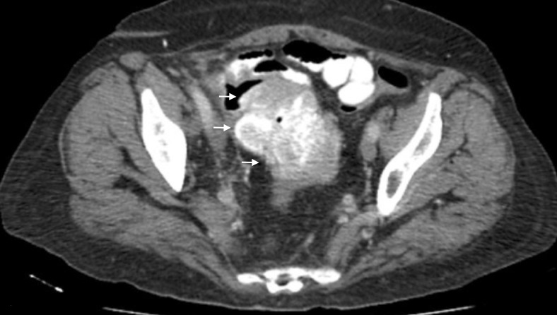
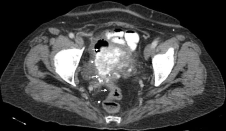
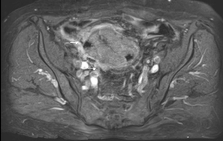
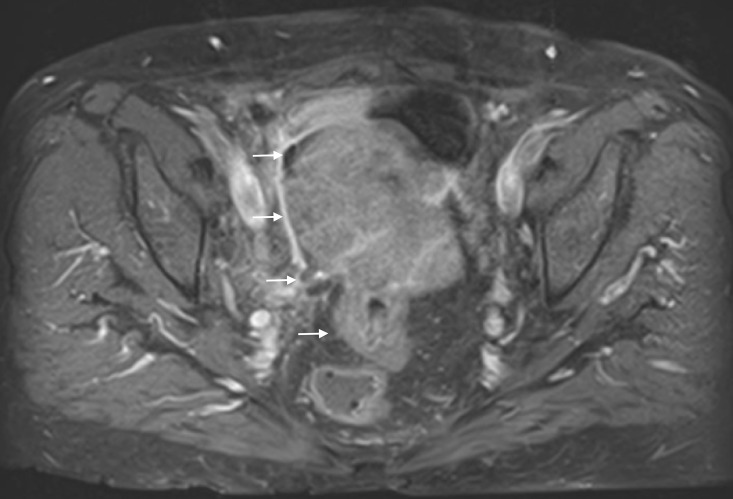
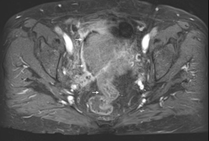
Myiasis is the infestation of humans with dipterous larvae. Traditionally, myiasis was thought to affect individuals living in tropical regions, however, several cases in temperate zones have been reported. We encountered two patients with histories of malignancies that presented with complaints of myiasis, in Chicago, in the spring and summer of 2016. The first patient, a 54-year-old female with a history of breast cancer, presented with complaints of maggots infesting her postsurgical chest wounds. She was diagnosed with sepsis, cellulitis, and wound myiasis. The second patient, a 63-year-old female with a history of recurrent ovarian cancer, presented with complaints of passing maggots vaginally and seeing worms mixed with her stools. She was diagnosed with internal urogenital myiasis. The first lesson that we learned from these cases is that myiasis can occur in individuals living in any part of the world. Second of all, for patients with accidental myiasis, a sample of the larvae should be sent for analysis to help guide the treatment. Third of all, myiasis has been associated with new or recurrent malignancies, and therefore a biopsy of the affected tissue should be sent for analysis. Finally, we learned that myiasis can serve as a form of tissue debridement; this coinciding benefit should not prevent the treatment of accidental myiasis.
Keywords: gynecological cancers; larvae; maggot therapy; malignant wound; myiasis.
Conflict of interest statement
The authors have declared that no competing interests exist.
Figures
References
-
- Tungiasis and myiasis. Cestari TF, Pessato S, Ramos-e-Silva M. Clin Dermatol. 2007;25:158–164. - PubMed
-
- Myiasis of the tracheostomy wound: case report. Franza R, Leo L, Minerva T, et al. https://www.ncbi.nlm.nih.gov/pmc/articles/PMC2639996/pdf/0392-100X.26.22.... Acta Otorhinolaryngol Ital. 2006;26:222–224. - PMC - PubMed
-
- Cutaneous myiasis. McGraw TA, Turiansky GW. J Am Acad Dermatol. 2008;58:907–926. - PubMed
-
- Myiasis in a case of invasive ductal carcinoma breast - a rare presentation. Kumar N, Nair RP, Sinha A, et al. http://medrech.com/sites/default/files/articles/53%20MYIASIS%20IN%20A%20... Med Res Chron. 2014;1:208–212.
Publication types
LinkOut - more resources
Full Text Sources
Other Literature Sources