

Trabecular bone microstructure is impaired in the proximal femur of human immunodeficiency virus-infected men with normal bone mineral density
- PMID: 29541618
- PMCID: PMC5835656
- DOI: 10.21037/qims.2017.10.10
Trabecular bone microstructure is impaired in the proximal femur of human immunodeficiency virus-infected men with normal bone mineral density
Abstract
Background: There is evidence that human immunodeficiency virus (HIV) infection and antiretroviral therapy (ART) are independent risk factors for osteoporosis and fracture which is not solely explained by changes in bone mineral density. Thus, we hypothesized that the assessment of trabecular microstructure might play an important role for bone quality in this population and might explain the increased fracture risk. In this study, we have assessed bone microstructure in the proximal femur using high-resolution magnetic resonance imaging (MRI) as well as in the extremities using high resolution peripheral quantitative computed tomography (HR-pQCT) in HIV-infected men and healthy controls and compared these findings to those based on areal bone mineral density (aBMD) derived from dual X-ray absorptiometry (DXA) which is the standard clinical parameter for the diagnosis of osteoporosis.
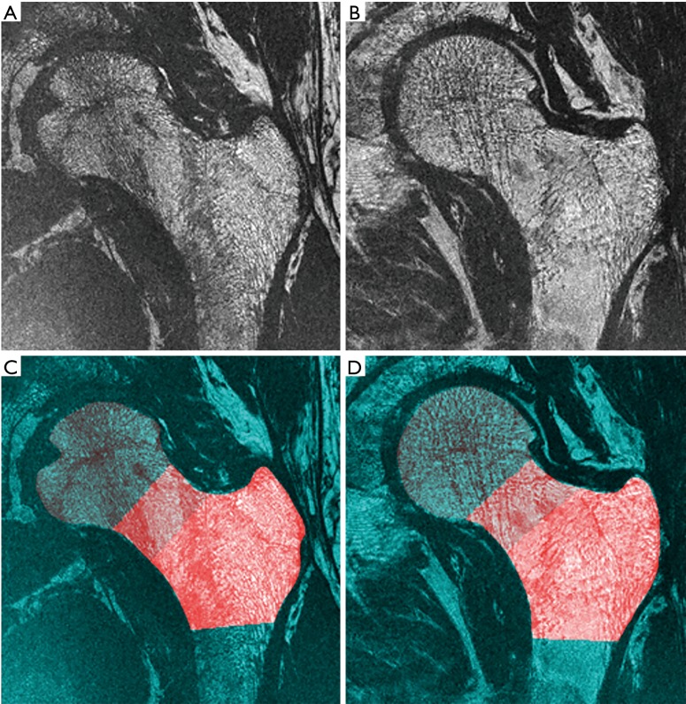
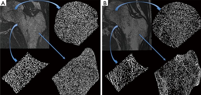
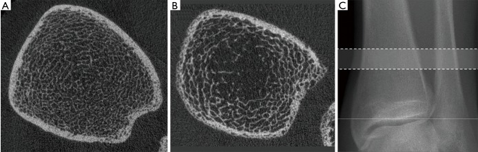
Methods: Eight HIV-infected men and 11 healthy age-matched controls were recruited and informed consent was obtained before each scan. High-resolution MRI of the proximal femur was performed using fully balanced steady state free precession (bSSFP) on a 3T system. Three volumes of interest at corresponding anatomic locations across all subjects were defined based on registrations of a common template. Four MR-based trabecular microstructural parameters were analyzed at each region: fuzzy bone volume fraction (f-BVF), trabecular number (Tb.N), thickness (Tb.Th), and spacing (Tb.Sp). In addition, the distal radius and distal tibia were imaged with HR-pQCT. Four HR-pQCT-based microstructural parameters were analyzed: trabecular bone volume fraction (BV/TV), Tb.N, Tb.Th, and Tb.Sp. Total hip and spine aBMD were determined from DXA.
Results: Microstructural bone parameters derived from MRI at the proximal femur and from HR-pQCT at the distal tibia showed significantly lower bone quality in HIV-infected patients compared to healthy controls. In contrast, DXA aBMD data showed no significant differences between HIV-infected patients and healthy controls.
Conclusions: Our results suggest that high-resolution imaging is a powerful tool to assess trabecular bone microstructure and can be used to assess bone health in HIV-infected men who show no differences to healthy males by DXA aBMD. Advances in MRI technology have made microstructural imaging at the proximal femur possible. Further studies in larger patient cohorts are clearly warranted.
Keywords: Human immunodeficiency virus (HIV); areal bone mineral density (aBMD); dual X-ray absorptiometry (DXA); high resolution peripheral quantitative computed tomography (HR-pQCT); high-resolution magnetic resonance imaging (MRI); trabecular bone microstructure.
Conflict of interest statement
Conflicts of Interest: The authors have no conflicts of interest to declare.
Figures
Similar articles
-
Bone microstructure in healthy men measured by HR-pQCT: Age-related changes and their relationships with DXA parameters and biochemical markers.Bone. 2022 Jan;154:116252. doi: 10.1016/j.bone.2021.116252. Epub 2021 Nov 4. Bone. 2022. PMID: 34743043
-
Longitudinal evaluation of the effects of alendronate on MRI bone microarchitecture in postmenopausal osteopenic women.Bone. 2011 Mar 1;48(3):611-21. doi: 10.1016/j.bone.2010.10.179. Epub 2010 Nov 5. Bone. 2011. PMID: 21059422 Free PMC article. Clinical Trial.
-
Automated simulation of areal bone mineral density assessment in the distal radius from high-resolution peripheral quantitative computed tomography.Osteoporos Int. 2009 Dec;20(12):2017-24. doi: 10.1007/s00198-009-0907-0. Epub 2009 Mar 28. Osteoporos Int. 2009. PMID: 19330422 Free PMC article.
-
MRI-Based Quantitative Osteoporosis Imaging at the Spine and Femur.J Magn Reson Imaging. 2021 Jul;54(1):12-35. doi: 10.1002/jmri.27260. Epub 2020 Jun 25. J Magn Reson Imaging. 2021. PMID: 32584496 Review.
-
[Review of high-resolution peripheral quantitative computed tomography for the assessment of bone microstructure and strength].Sheng Wu Yi Xue Gong Cheng Xue Za Zhi. 2018 Jun 25;35(3):468-474. doi: 10.7507/1001-5515.201707068. Sheng Wu Yi Xue Gong Cheng Xue Za Zhi. 2018. PMID: 29938957 Free PMC article. Review. Chinese.
Cited by
-
Evaluation of bone mineral density, microarchitecture, and detection of fractures on young patients living with human immunodeficiency virus: when and how to screen?Endocrine. 2024 Jan;83(1):214-226. doi: 10.1007/s12020-023-03501-9. Epub 2023 Sep 6. Endocrine. 2024. PMID: 37673836
-
Survey of MRI Usefulness for the Clinical Assessment of Bone Microstructure.Int J Mol Sci. 2021 Mar 2;22(5):2509. doi: 10.3390/ijms22052509. Int J Mol Sci. 2021. PMID: 33801539 Free PMC article. Review.
-
People living with HIV have low trabecular bone mineral density, high bone marrow adiposity, and poor trabecular bone microarchitecture at the proximal femur.Osteoporos Int. 2022 Aug;33(8):1739-1753. doi: 10.1007/s00198-022-06405-y. Epub 2022 Apr 27. Osteoporos Int. 2022. PMID: 35478045 Free PMC article.
-
Investigation of Osteoporosis in Persons Living with Human Immunodeficiency Virus: The HOST Study.Calcif Tissue Int. 2025 Apr 25;116(1):64. doi: 10.1007/s00223-025-01368-8. Calcif Tissue Int. 2025. PMID: 40281238 Free PMC article.
-
Long Bone Mineral Loss, Bone Microstructural Changes and Oxidative Stress After Eimeria Challenge in Broilers.Front Physiol. 2022 Jul 18;13:945740. doi: 10.3389/fphys.2022.945740. eCollection 2022. Front Physiol. 2022. PMID: 35923236 Free PMC article.
References
Grants and funding
LinkOut - more resources
Full Text Sources
Other Literature Sources