Review
. 2018 Feb;8(1):100-113.
doi: 10.21037/qims.2018.02.02.

Clinical utilities of quantitative ultrasound in osteoporosis associated with inflammatory rheumatic diseases

Affiliations
Review

Clinical utilities of quantitative ultrasound in osteoporosis associated with inflammatory rheumatic diseases

Win Min Oo et al. Quant Imaging Med Surg. 2018 Feb.

Abstract

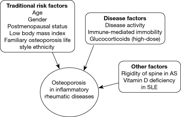
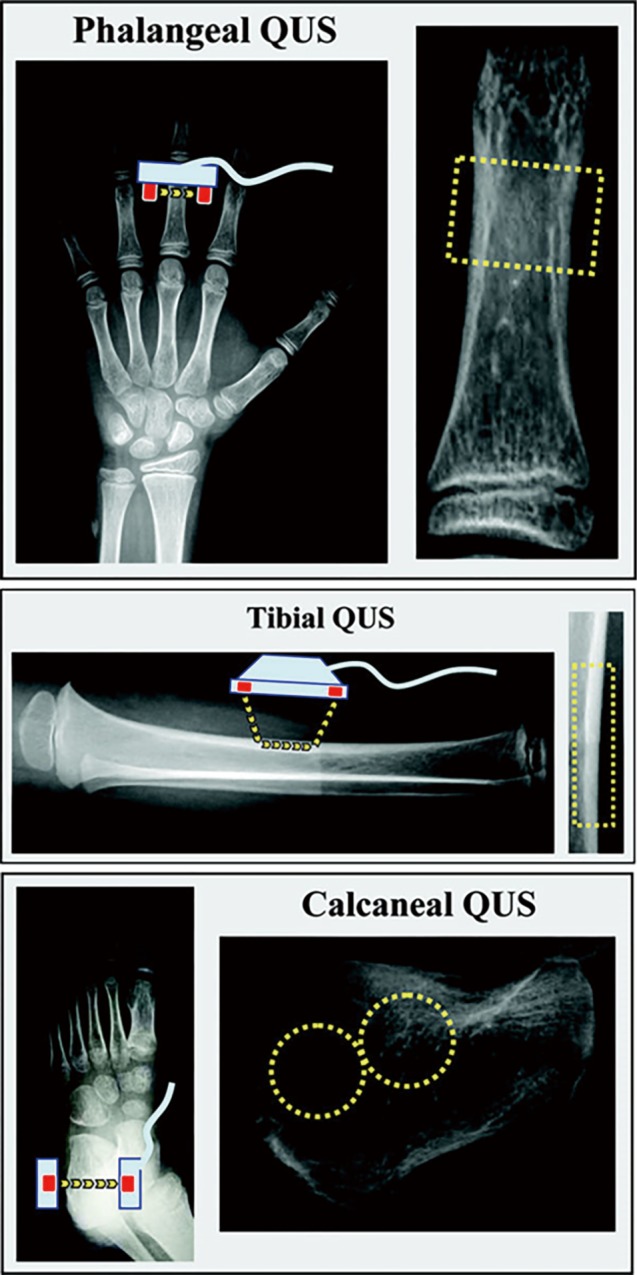
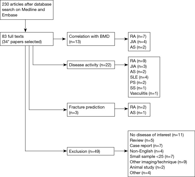
Secondary osteoporosis is an important co-morbidity related to inflammatory rheumatic diseases that is attributed to several factors including inflammatory cytokines, inactivity and glucocorticoid treatment. Quantitative ultrasound (QUS) has been utilized in osteoporosis research due to its detectability of bone density as well as bone quality. The current narrative review is to address the potential utilities of QUS in secondary osteoporosis of inflammatory rheumatic diseases, focusing on the clinical aspects of QUS in these diseases, based on the conformity of QUS with dual emission X-ray absorptiometry (DXA), the relationship with disease characteristics, and its capability of fracture prediction. Although limited data demonstrate that QUS had moderate to strong correlation with DXA, and might be useful as a potential imaging tool to screen for osteoporosis, further research is still required for QUS to be utilized effectively for the best outcome in these patients with rheumatic diseases.

Keywords: Quantitative ultrasound (QUS); fracture; inflammatory rheumatic disease; osteoporosis; review.


Conflict of interest statement

Conflicts of Interest: The authors have no conflicts of interest to declare.

Figures

Figure 1
Figure 1
Risk factors for osteoporosis and fractures in inflammatory rheumatic diseases. AS, ankylosing spondylitis; SLE, systemic lupus erythematosus.
Figure 2
Figure 2
Areal BMD measurements are influenced by bone size, with larger bones of identical volumetric densities having higher areal BMD values. Reproduced from (43) (reprinted by permission from the American Society for Bone and Mineral Research). BMD, bone mineral density.
Figure 3
Figure 3
Quantitative computed tomography measuring osteoporosis of lumber spine. (A) For three-dimensional volumetric quantitative computed tomography protocols, a contiguous volume with a slice thickness of 1–3 mm with no computed tomography gantry tilt is typically scanned; (B) at the spine, a region of interest marked in red circle measures the trabecular bone mineral density in isolation (52). (Courtesy J. Keenan Brown, Mindways Software Inc., Austin, TX, USA).
Figure 4
Figure 4
Analysis of the trace of the sound wave to calculate SoS, AD-SoS and BTT (62). (Adapted by permission from Macmillan Publishers Ltd.). SoS, speed of sound; AD-SoS, amplitude-dependent speed of sound.
Figure 5
Figure 5
Schematic representation of QUS devices (phalangeal, tibial and calcaneal) used to assess bone mineral status. X-ray films are used to indicate the skeletal site of measurement, and the approximate ROI is depicted on the right side for each device. The probe(s) is in light blue and the transducers are in red; the yellow arrows show the principal pathways of the ultrasound waves from the emitter transducer(s) to the receiving transducer(s). The yellow rectangle represents the ROI for phalangeal and tibial devices while the yellow rings show the approximate ROI for two different calcaneal QUS devices (62). (Reprinted with permission from Macmillan Publishers Ltd.). QUS, quantitative ultrasound.
Figure 6
Figure 6
Flow diagram of literature search. *, several papers examined more than one QUS utility. BMD, bone mineral density; RA, rheumatoid arthritis; JIA, juvenile idiopathic arthritis; AS, ankylosing spondylitis; SLE, systemic lupus erythematosus; PS, psoriatic arthritis; SS, systemic sclerosis.

References

    1. Consensus development conference : diagnosis, prophylaxis, and treatment of osteoporosis. Am J Med 1993;94:646-50. 10.1016/0002-9343(93)90218-E - DOI - PubMed
    1. Assessment of fracture risk and its application to screening for postmenopausal osteoporosis. Report of a WHO Study Group. World Health Organ Tech Rep Ser 1994;843:1-129. - PubMed
    1. Gordon CM, Baim S, Bianchi ML, Bishop NJ, Hans DB, Kalkwarf H, Langman C, Leonard MB, Plotkin H, Rauch F, Zemel BS. Special report on the 2007 Pediatric Position Development Conference of the International Society for Clinical Densitometry. South Med J 2008;101:740-3. 10.1097/SMJ.0b013e31817a8b55 - DOI - PubMed
    1. Binkley TL, Berry R, Specker BL. Methods for measurement of pediatric bone. Rev Endocr Metab Disord 2008;9:95-106. 10.1007/s11154-008-9073-5 - DOI - PubMed
    1. Ekman EF. The role of the orthopaedic surgeon in minimizing mortality and morbidity associated with fragility fractures. J Am Acad Orthop Surg 2010;18:278-85. 10.5435/00124635-201005000-00004 - DOI - PubMed

LinkOut - more resources