Review
. 2018 Feb 28:6:17.
doi: 10.3389/fcell.2018.00017. eCollection 2018.

Feeling Stress: The Mechanics of Cancer Progression and Aggression

Affiliations
Review

Feeling Stress: The Mechanics of Cancer Progression and Aggression

Josette M Northcott et al. Front Cell Dev Biol. .

Abstract

The tumor microenvironment is a dynamic landscape in which the physical and mechanical properties evolve dramatically throughout cancer progression. These changes are driven by enhanced tumor cell contractility and expansion of the growing tumor mass, as well as through alterations to the material properties of the surrounding extracellular matrix (ECM). Consequently, tumor cells are exposed to a number of different mechanical inputs including cell-cell and cell-ECM tension, compression stress, interstitial fluid pressure and shear stress. Oncogenes engage signaling pathways that are activated in response to mechanical stress, thereby reworking the cell's intrinsic response to exogenous mechanical stimuli, enhancing intracellular tension via elevated actomyosin contraction, and influencing ECM stiffness and tissue morphology. In addition to altering their intracellular tension and remodeling the microenvironment, cells actively respond to these mechanical perturbations phenotypically through modification of gene expression. Herein, we present a description of the physical changes that promote tumor progression and aggression, discuss their interrelationship and highlight emerging therapeutic strategies to alleviate the mechanical stresses driving cancer to malignancy.

Keywords: ECM stiffness; cancer progression; cell contractility; mechanical stresses; solid stress; therapeutic targets; tissue tension.


Figures
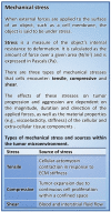
Figure 1
Figure 1
Mechanical stress.
Figure 2
Figure 2
Mechanical forces and cellular contractility. (A) Forces exerted onto a cell can cause three types of mechanical stress: tensile (stretch), compressive, and shear. Forces that induce tensile and compressive stress are applied perpendicular to the cell surface, while forces causing shear stress are exerted parallel to the cell surface. (B) Integrin clusters activated through ECM binding are bound by talin, initiating actin polymerization and producing intracellular tension. In response to high tensile force (a consequence of elevated ECM rigidity), FAK is activated (top). Activation of FAK leads to the recruitment of additional linker proteins, focal adhesion maturation, and increased actomyosin contraction via the assembled actin stress fibers (bottom).
Figure 3
Figure 3
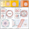
Diverse mechanical stimuli act on tumor cells throughout cancer progression. Simplified depiction of oncogenic transformation and solid tumor progression for cancers of epithelial origin (top). Letters indicate the stages of cancer progression focused on in panels (A–D). (A) In homeostatic tissues, the forces between cells and the ECM are balanced. (B) The tumor microenvironment is composed of cancer cells with augmented contractility (increased intracellular tension), surrounded by a progressively stiffening ECM (increased ECM resistance), and a host of stromal cell types including fibroblasts, immune cells and vascular cell types. (C) Tumor expansion confined by the surrounding stroma compresses both the tumor and the adjacent stromal tissue, causing increased interstitial pressure. Augmented ECM rigidity increases stromal resistance to compression and exacerbates solid stress. (D) A high interstitial fluid pressure gradient elicits fluid flow from the tumor core to the periphery, promoting metastatic dissemination. Following escape from the primary tumor, cancer cells migrate along tension-oriented collagen fibers toward the vasculature. Tumor cells are exposed to high shear stresses as they intravasate/extravasate between endothelial cells and travel through the circulation en route to future secondary tumor sites.
Figure 4
Figure 4
Relationships between mechanical stress and tissue responses during tumor progression to metastasis. Increased intracellular tension is produced through actomyosin contraction in response to both biochemical and mechanical stimuli. ECM stiffening stimulates cells to generate higher intracellular tension to exert stronger traction forces on their surroundings, which subsequently exacerbates ECM stiffness. Solid stress is caused in part by unchecked proliferation of cancer cells that results in expansion of the tumor mass, compression of the tumor interior and distention of the surrounding stromal tissue. ECM stiffness can increase solid stress by augmenting the resistance to tumor expansion. Reciprocally, tumor expansion causes circumferential ECM tension and tissue stiffening. As a result of tissue compression and ECM stiffening, blood and lymphatic vascular function is impaired (due to vascular crushing) and interstitial fluid pressure is increased. Aberrant fluid flow throughout the interstitial spaces and within the obstructed tumor vasculature increases the shear stress experienced by tumor cells.

Similar articles

Cited by

References

    1. Acerbi I., Cassereau L., Dean I., Shi Q., Au A., Park C., et al. . (2015). Human breast cancer invasion and aggression correlates with ECM stiffening and immune cell infiltration. Integr. Biol. 7, 1120–1134. 10.1039/C5IB00040H - DOI - PMC - PubMed
    1. Aung A., Seo Y. N., Lu S., Wang Y., Jamora C., del Álamo J. C., et al. . (2014). 3D traction stresses activate protease-dependent invasion of cancer cells. Biophys. J. 107, 2528–2537. 10.1016/j.bpj.2014.07.078 - DOI - PMC - PubMed
    1. Avvisato C. L., Yang X., Shah S., Hoxter B., Li W., Gaynor R., et al. . (2007). Mechanical force modulates global gene expression and beta-catenin signaling in colon cancer cells. J. Cell Sci. 120(Pt 15), 2672–2682. 10.1242/jcs.03476 - DOI - PubMed
    1. Basson M. D., Zeng B., Downey C., Sirivelu M. P., Tepe J. J. (2015). Increased extracellular pressure stimulates tumor proliferation by a mechanosensitive calcium channel and PKC-beta. Mol. Oncol. 9, 513–526. 10.1016/j.molonc.2014.10.008 - DOI - PMC - PubMed
    1. Basu R., Whitlock B. M., Husson J., Le Floc'h A., Jin W., Oyler-Yaniv A., et al. . (2016). Cytotoxic T cells use mechanical force to potentiate target cell killing. Cell 165, 100–110. 10.1016/j.cell.2016.01.021 - DOI - PMC - PubMed