. 2018 Feb 10:10:96-100.
doi: 10.1016/j.ajoc.2018.02.008. eCollection 2018 Jun.

Acute macular neuroretinopathy associated with influenza vaccination with decreased flow at the deep capillary plexus on OCT angiography

Affiliations

Acute macular neuroretinopathy associated with influenza vaccination with decreased flow at the deep capillary plexus on OCT angiography

Jeffrey C Liu et al. Am J Ophthalmol Case Rep. .

Abstract

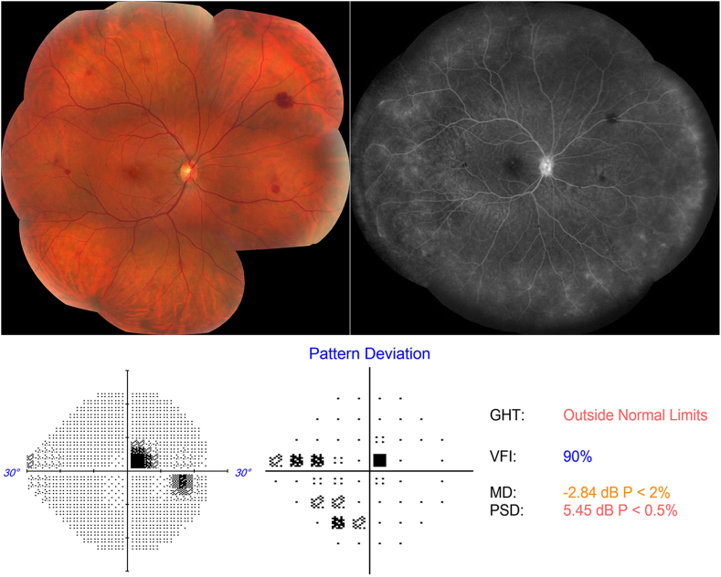
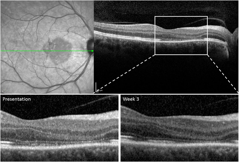
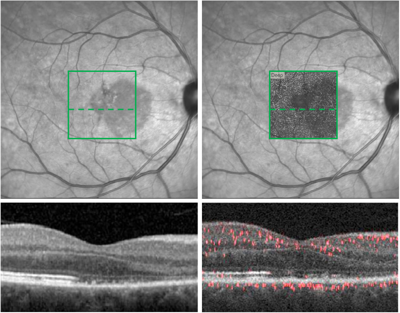
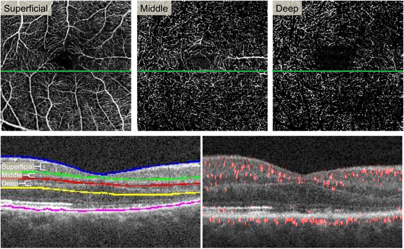
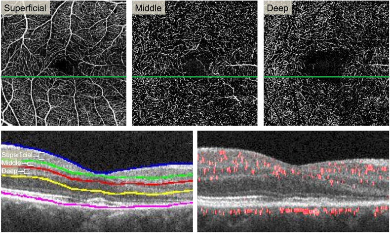
Purpose: We report a case of acute macular neuroretinopathy (AMN) following routine annual inactivated influenza vaccination. Projection-resolved optical coherence tomography angiography (PR-OCTA) was used to analyze the retinal capillary flow within the AMN lesion.

Observations: Our patient reported visual symptoms of her right eye nine days after routine annual influenza vaccination. Multimodal imaging revealed small vessel peripheral vasculitis and AMN in the affected eye. Infectious, immunologic, and hypercoagulable etiologies were investigated and excluded. PR-OCTA B-scans within the AMN lesion demonstrated reduced flow in the deep capillary plexus (DCP) at baseline with relatively improved flow signal in the DCP on follow up, 3 weeks later.

Conclusions and importance: We report a new association of AMN following routine inactivated influenza immunization. Recent influenza vaccination should be included in the differential diagnosis for patients presenting with AMN. PR-OCTA demonstrated compromised DCP flow in the AMN lesion which has not been previously described.

Keywords: AMN; Acute macular neuroretinopathy; Influenza vaccine; OCTA; Optical coherence tomography angiography; Vasculitis.


Figures

Fig. 1
Fig. 1
Color fundus photo, fluorescein angiogram (FA) and Humphrey 242 Threshold perimetry on presentation. Left: Color fundus photo montage of the right eye shows macular pigment mottling and mid-peripheral intra-retinal blot hemorrhages. Right: late FA shows staining of the disc and diffuse leakage and staining of the peripheral capillary vasculature. Bottom: Demonstrates a right superotemporal paracentral scotoma. (For interpretation of the references to colour in this figure legend, the reader is referred to the Web version of this article.)
Fig. 2
Fig. 2
Spectral Domain (SD-OCT) within the acute macular neuroretinopathy (AMN) lesion on presentation and week 3. Top Left: On presentation, near-Infrared imaging shows wedge-shaped hypo-reflective macular lesion. Top Right, Bottom Left: SD-OCT shows thinning of the outer nuclear layer and disruption of the ellipsoid and interdigitation zones. Bottom Right: 3 weeks later, SD-OCT shows persistent thinning of outer nuclear layer with some restoration of the ellipsoid and interdigitation zones.
Fig. 3
Fig. 3
Spectral Domain (SD-OCT) and Projection-Resolved (PR-OCTA) overlay within the acute macular neuroretinopathy (AMN) lesion at presentation. Top Left: Near-Infrared image with represented 3 × 3mm2en face PR-OCTA area (green square) and corresponding cross-sectional segment (dashed green line). Bottom left: Cross-sectional SD-OCT within the AMN lesion in the represented 3 × 3mm2 area. Top Right: Near-Infrared image with 3 × 3mm2en face PR-OCTA deep capillary plexus (DCP) slab overlay and corresponding cross-sectional segment (dashed green line). Bottom Right: Cross-sectional PR-OCTA within the AMN lesion. (For interpretation of the references to colour in this figure legend, the reader is referred to the web version of this article.)
Fig. 4
Fig. 4
Projection-resolved (PR-OCTA) reveals attenuated flow signal in the deep capillary plexus. Top Row:En face PR-OCTA projections of the superficial, middle, and deep capillary plexuses. Some large superficial vessels are seen in the middle plexus due to segmentation error. The deep plexus shows signal attenuation in the areas of the lesion (far right). Bottom Left: Cross-sectional SD-OCT showing segmentation lines for the three plexuses. Bottom Right: Cross-sectional PR-OCTA with red flow overlay reveals reduced red flow signal in the inner nuclear layer and outer plexiform layer in the area of the lesion (right half). (For interpretation of the references to colour in this figure legend, the reader is referred to the web version of this article.)
Fig. 5
Fig. 5
Projection-resolved (PR-OCTA) on follow-up shows increased flow signal in the deep capillary plexus. Top Row:En face PR-OCTA projections of the superficial, middle, and deep capillary plexuses. Some large superficial vessels are seen in the middle plexus due to segmentation error (center). The deep plexus shows some signal attenuation in the areas of the lesion (far right), but more flow compared to presentation (Fig. 4). Bottom Left: Cross-sectional SD-OCT showing segmentation lines for the three plexuses. Bottom Right: Cross-sectional PR-OCTA with red flow overlay reveals some recovery of flow signal in the inner nuclear layer and outer plexiform layer in the area of the lesion (right half).

References

    1. Bos P.J., Deutman A.F. Acute macular neuroretinopathy. Am J Ophthalmol. 1975;80(4):573–584. - PubMed
    1. Hurwitz E.S., Schonberger L.B., Nelson D.B., Holman R.C. Guillain-Barré syndrome and the 1978-1979 influenza vaccine. N Engl J Med. 1981;304(26):1557–1561. - PubMed
    1. Carrillo-santisteve P., Ciancio B.C., Nicoll A., Lopalco P.L. The importance of influenza prevention for public health. Hum Vaccin Immunother. 2012;8(1):89–95. - PubMed
    1. Grohskopf L.A., Olsen S.J., Sokolow L.Z. Prevention and control of seasonal influenza with vaccines: recommendations of the Advisory Committee on Immunization Practices (ACIP) – United States, 2014-15 influenza season. MMWR Morb Mortal Wkly Rep. 2014;63(32):691–697. - PMC - PubMed
    1. Williams G.S., Evans S., Yeo D., Al-bermani A. Retinal artery vasculitis secondary to administration of influenza vaccine. BMJ Case Rep. 2015 - PMC - PubMed