Review
. 2018 Apr;9(2):215-236.
doi: 10.1007/s13244-018-0597-2. Epub 2018 Mar 14.

Vascular CT and MRI: a practical guide to imaging protocols

Affiliations
Review

Vascular CT and MRI: a practical guide to imaging protocols

D J Murphy et al. Insights Imaging. 2018 Apr.

Abstract

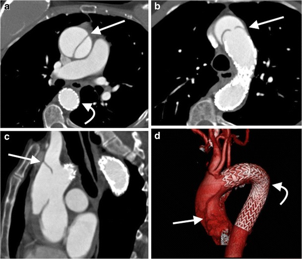
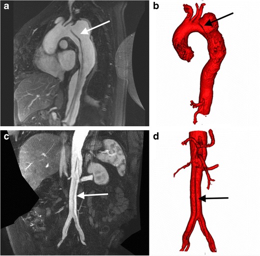
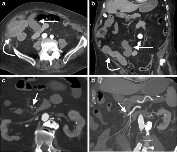
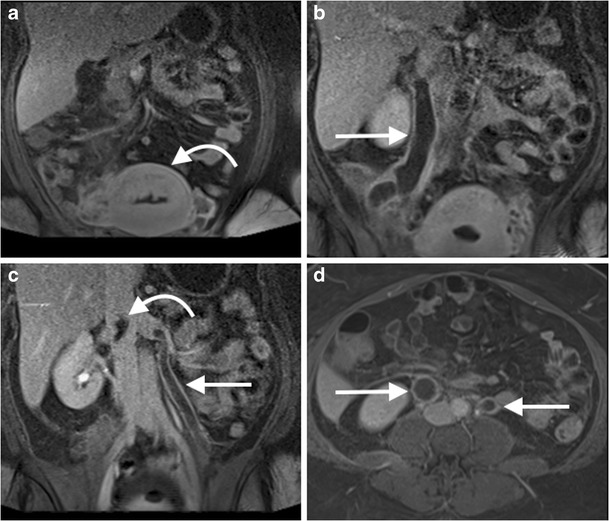
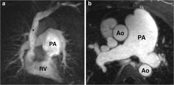
Non-invasive cross-sectional imaging techniques play a crucial role in the assessment of the varied manifestations of vascular disease. Vascular imaging encompasses a wide variety of pathology. Designing vascular imaging protocols can be challenging owing to the non-uniform velocity of blood in the aorta, differences in cardiac output between patients, and the effect of different disease states on blood flow. In this review, we provide the rationale behind-and a practical guide to-designing and implementing straightforward vascular computed tomography (CT) and magnetic resonance imaging (MRI) protocols.Teaching Points • There is a wide range of vascular pathologies requiring bespoke imaging protocols. • Variations in cardiac output and non-uniform blood velocity complicate vascular imaging. • Contrast media dose, injection rate and duration affect arterial enhancement in CTA. • Iterative CT reconstruction can improve image quality and reduce radiation dose. • MRA is of particular value when imaging small arteries and venous studies.

Keywords: Angiography; Atherosclerosis; Computed tomography angiography; Magnetic resonance angiography; Magnetic resonance imaging.


Figures

Fig. 1
Fig. 1
A 45-year-old man with Marfan’s syndrome post aortic valve replacement with a fusiform ascending aortic aneurysm. Measurement of ascending thoracic aorta dimension (double arrows) in an axial plane (a) will yield erroneous values due to oblique orientation relative to the centerline of the aorta, as demonstrated on sagittal (red line, b) and coronal (red line, c) multiplanar reformats (MPRs). Three-dimensional segmented volume rendered (VR) image of the thoracic aorta (d) demonstrates the site of measurement (line)
Fig. 2
Fig. 2
A 45-year-old man with Marfan’s syndrome post aortic valve replacement with a fusiform ascending aortic aneurysm. Measurement of the ascending thoracic aorta dimension (double arrows) in a plane double oblique to the vessel centerline (a) with sagittal (b) and coronal (c) MPRs demonstrating the plane of measurement (red lines). Three-dimensional segmented VR image of the thoracic aorta (d) demonstrates the site of measurement (line)
Fig. 3
Fig. 3
a Three-dimensional segmented volume rendered (VR) image of the thoracic aorta with b standard sites of measurement. a Sinuses of Valsalva, b sinotubular junction, c ascending thoracic aorta, d aortic arch, between origin of left common carotid and left subclavian arteries, e descending thoracic aorta, f aortic hiatus
Fig. 4
Fig. 4
a Three-dimensional segmented VR image of the abdominal aorta with b standard sites of measurement. a Proximal abdominal aorta, b juxtarenal abdominal aorta, c infrarenal abdominal aorta, d right common iliac artery, e left common iliac artery
Fig. 5
Fig. 5
Axial (a, b) and sagittal (c) arterial phase images from an ECG-gated CT thoracic aorta angiogram in a 62-year-old woman with chest pain demonstrates a type A dissection in the ascending thoracic aorta (a-c, arrow) with evidence of prior stent graft repair of the descending thoracic aorta (a-c, curved arrow). Three-dimensional segmented VR image of the thoracic aorta (d) delineates the ascending aorta dissection flap (arrow) and descending thoracic aorta stent graft (curved arrow)
Fig. 6
Fig. 6
A MR angiogram in a 45-year-old woman who presented with acute onset of tearing chest pain. Sagittal oblique image from a ECG gated T1-weighted post-gadolinium 3D acquisition (a) demonstrates a dissection flap (arrow) arising in the aortic arch distal to the origin of left subclavian artery consistent with a type B aortic dissection, with the dissection flap extending into the abdominal aorta. Corresponding 3D segmented volume rendered image of the thoracic aorta (b) from the thoracic MRA demonstrates the dissection flap (arrow). Coronal abdominal T1-weighted post gadolinium MRA image (c) demonstrates the distal extent of the dissection flap (arrow) into the common iliac arteries bilaterally, with a 3D segmented volume rendered image of the abdominal aorta (d) demonstrating the dissection flap in the abdominal aorta (arrow)
Fig. 7
Fig. 7
Selected axial images from T1-weighted 3D spoiled gradient echo sequence with a fat selective prepulse sequence pre- (a) and post- (b) gadolinium in a 55-year-old woman with giant cell arteritis demonstrates enhancing circumferential mural aortic soft thickening (arrow) of the juxtarenal aorta (Ao) consistent with a large vessel vasculitis
Fig. 8
Fig. 8
Selected images from a CT angiogram in a 55-year-old man undergoing imaging surveillance post abdominal aortic aneurysm endovascular stent graft repair (EVAR). Axial non-contrast (a), arterial phase (b) and 70 s delayed phase (c) images demonstrate iodinated contrast material within the excluded aneurysm sac (arrow) consistent with an endoleak. Sagittal oblique MPR of the right common iliac artery (d) demonstrates the endoleak arising from the distal insertion point of the right iliac limb of the EVAR consistent with a type 1b endoleak
Fig. 9
Fig. 9
Selected axial images (a-d) from an arterial phase CT angiogram of the abdominal aorta in a 55-year-old man with hypotension and abdominal pain demonstrates a large retroperitoneal haematoma (curved arrow) and active extravasation from the infrarenal abdominal aorta (arrow) consistent with aortic rupture. A ruptured mycotic aneurysm was found in the operating room
Fig. 10
Fig. 10
A 50-year-old man with oesophageal cancer was referred to CT following discovery of a mesenteric haematoma during exploratory laparoscopy. Axial non-contrast (a), arterial phase (b) and 70-s post contrast (c) images demonstrate an ill-defined mesenteric haematoma (arrow), a pancreaticoduodenal arcade pseudoaneurysm (curved arrow), with no active extravasation. The patient was taken to the interventional radiology suite and the pseudoaneurysm was occluded with coils; selected spot fluoroscopic image (d) demonstrates the occluded pancreaticoduodenal arcade filled with coils (curved arrow)
Fig. 11
Fig. 11
An 85-year-old woman developed severe abdominal pain two days post percutaneous aortic valve replacement and was referred for a CT mesenteric angiogram. Axial (a) and coronal (b) images from an arterial phase CT mesenteric angiogram demonstrate hypoenhancement of distal ileal loops (straight arrow) compared with adjacent proximal ileal and jejunal loops with normal mural enhancement (curved arrow). Axial (c) and coronal oblique images from the same study demonstrate focal occlusion of the mid-superior mesenteric artery with calcified plaque (arrow). Coronal oblique maximum intensity projection (MIP) reformat (d) is useful in demonstrating the point of SMA obstruction (arrow). Exploratory laparoscopy revealed extensive small bowel ischaemia, and the patient unfortunately expired
Fig. 12
Fig. 12
Three dimensional segmented volume rendered image of the kidneys and their arterial supply in a renal donor volunteer, segmented from an arterial phase renal artery angiogram CT, demonstrates bilateral accessory renal arteries supplying the lower renal poles
Fig. 13
Fig. 13
A 45-year-old man with bilateral leg swelling underwent an MR venogram of the abdomen and pelvis. Coronal image from a T2-weighted sequence (a) demonstrated an expanded suprarenal IVC with heterogeneous T2 high signal material, likely thrombus (arrow). Coronal (b) and axial (c) images from a venous phase T1-weighted fat-saturated post-gadolinium sequence demonstrates heterogeneous enhancement of the intraluminal IVC material, concerning for tumour thrombus. A coronal image from a T1-weighted fat-saturated post-gadolinium sequence of the abdomen (d) demonstrates a horseshoe kidney with an exophytic mass arising from the right lower moiety (curved arrow), which was subsequently confirmed as a papillary renal cell carcinoma on biopsy
Fig. 14
Fig. 14
A 35-year-old woman underwent an abdominal MR venogram 5 days post dilatation and curettage for an intrauterine fetal death at 25 weeks. Coronal images from a post-contrast venous phase fat saturated T1 sequence of the abdomen demonstrates the post-partum uterus (a, curved arrow), with marked dilatation of the right (b, arrow) and left (c, arrow) gonadal veins with central filling defects consistent with bilateral gonadal vein thrombosis. The thrombus extends up to the juxtarenal IVC (c, curved arrow). Axial image from a T1 post-contrast venous phase fat saturated T1 sequence of the abdomen (d) demonstrates the expanded gonadal veins bilaterally with central filling defects
Fig. 15
Fig. 15
Selected sagittal images from a time-resolved MRA of the left foot in a patient with diabetes and foot pain. The initial mask image (a) demonstrates only a vague outline of the foot, with subsequent enhancement of the arteries (b, c), followed by venous filling at a later time point (d). This study demonstrates occlusion of the left dorsalis pedis in the left mid-foot (c, arrow), the likely cause of the patient symptoms
Fig. 16
Fig. 16
A 45-year-old man with a previous history of left radial artery thrombosis post coeliac artery stenting with persistent arm claudication. Time resolved MRA (a-d) of the left forearm demonstrates opacification of the left brachial artery, and left ulnar artery (arrows) with occlusion of the proximal left radial artery (curved arrows) (b, c), followed by venous filling (d)
Fig. 17
Fig. 17
A pulmonary artery MRA in a 55-year-old woman with pulmonary hypertension. Single coronal image from time-resolved pulmonary artery MRA (a) and axial T1-weighted fat-saturated post-gadolinium angiogram image (b) demonstrate fusiform aneurysmal dilatation of the main pulmonary artery (PA)
Fig. 18
Fig. 18
A 45-year-old woman with breast cancer undergoes a CT deep inferior epigastric artery perforator (DIEP) protocol prior to breast reconstruction. Coronal oblique 3D segmented VR image from the CT displays the abdominal wall musculature and superficial arterial supply. The location of the largest DIEP relative to the umbilicus is annotated with craniocaudal and mediolateral distance measurements to help the surgeon locate the vessel during surgery

References

    1. Aghayev A, Murphy DJ, Keraliya AR, Steigner ML. Recent developments in the use of computed tomography scanners in coronary artery imaging. Expert Rev Med Devices. 2016;6:545–553. doi: 10.1080/17434440.2016.1184968. - DOI - PubMed
    1. Otero HJ, Steigner ML, Rybicki FJ. The “post-64” era of coronary CT angiography: understanding new technology from physical principles. Radiol Clin N Am. 2009;47(1):79–90. doi: 10.1016/j.rcl.2008.11.001. - DOI - PMC - PubMed
    1. Flohr TG, De Cecco CN, Schmidt B, et al. Computed tomographic assessment of coronary artery disease: state-of-the-art imaging techniques. Radiol Clin N Am. 2015;53:271–285. doi: 10.1016/j.rcl.2014.11.011. - DOI - PubMed
    1. Kalisz K, Buethe J, Saboo SS, et al. Artifacts at cardiac CT: physics and solutions. Radiographics. 2016;36:2064–2083. doi: 10.1148/rg.2016160079. - DOI - PubMed
    1. Bae KT. Intravenous contrast medium administration and scan timing at CT: considerations and approaches. Radiology. 2010;256:32–61. doi: 10.1148/radiol.10090908. - DOI - PubMed

LinkOut - more resources