. 2018 Sep;23(5):813-826.
doi: 10.1007/s12192-018-0890-5. Epub 2018 Mar 14.

Small heat shock protein speciation: novel non-canonical 44 kDa HspB5-related protein species in rat and human tissues

Affiliations

Small heat shock protein speciation: novel non-canonical 44 kDa HspB5-related protein species in rat and human tissues

Rainer Benndorf et al. Cell Stress Chaperones. 2018 Sep.

Abstract

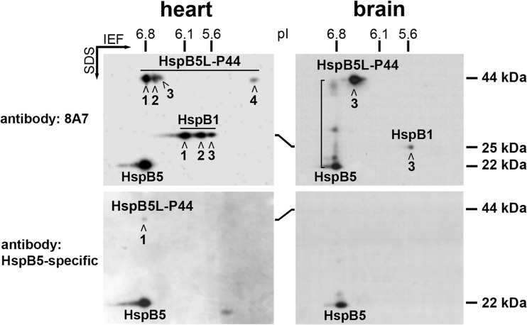
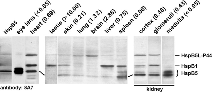
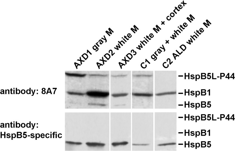
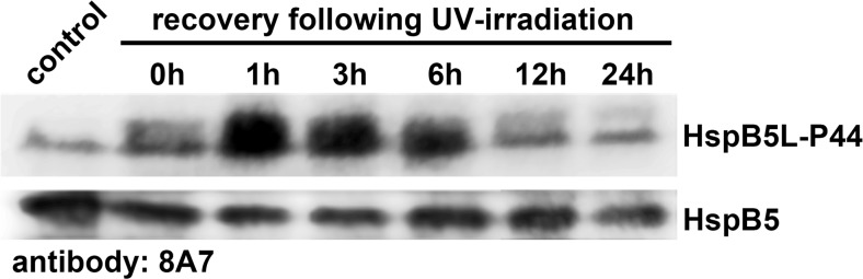
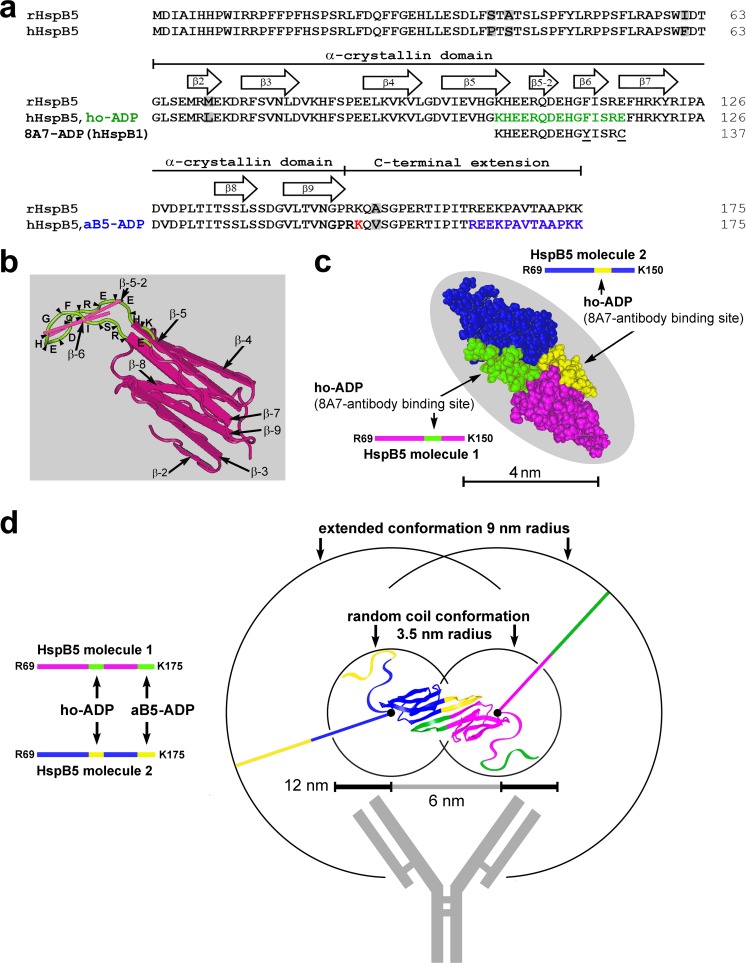
When analyzing small stress proteins of rat and human tissues by electrophoretic methods followed by western blotting, and using the anti-HspB1/anti-HspB5 antibody clone 8A7, we unexpectedly found a protein with a molecular mass of ~44 kDa. On two-dimensional gels, this protein resolved into four distinct species. Electrophoretic and immunological evidence suggests that this 44 kDa protein is a derivative of HspB5, most likely a covalently linked HspB5 dimer. This HspB5-like 44 kDa protein (HspB5L-P44) is particularly abundant in rat heart, brain, and renal cortex and glomeruli. HspB5L-P44 was also found in human brains, including those from patients with Alexander disease, a condition distinguished by cerebral accumulation of HspB5. Gray matter of such a patient contained an elevated amount of HspB5L-P44. A spatial model of structurally ordered dimeric HspB5 α-crystallin domains reveals the exposed and adjacent position of the two peptide segments homologous to the HspB1-derived 8A7 antigen determinant peptide (epitope). This explains the observed extraordinary high avidity of the 8A7 antibody towards HspB5L-P44, as opposed to commonly used HspB5-specific antibodies which recognize other epitopes. This scenario also explains the remarkable fact that no previous study reported the existence of HspB5L-P44 species. Exposure of rat endothelial cells to UV light, an oxidative stress condition, temporarily increased HspB5L-P44, suggesting physiological regulation of the dimerization. The existence of HspB5L-P44 supports the protein speciation discourse and fits to the concept of the protein code, according to which the expression of a given gene is reflected only by the complete set of the derived protein species.

Keywords: Covalently bonded HspB5 dimers; HspB1-/HspB5-specific antibody clone 8A7; Mammals; Protein modification; Protein speciation.


Conflict of interest statement

Rats were housed in animal facilities accredited by the American Association of Laboratory Animal Care, with free access to pelleted food and water. Housing and all procedures were conducted in accordance with the guidelines of the National Institute of Health and were approved by the University of Michigan Committee on Use and Care of Animals (approval nos. 7835 and 7989). Rats were euthanized by inhalation of carbon dioxide in accordance with the American Veterinary Medical Association guidelines on euthanasia.

Figures

Fig. 1
Fig. 1
Immunologic and 2D-PAGE evidence for an HspB5-related 44 kDa protein (HspB5L-P44) in rat heart and brain. The western blots were stained either with the 8A7 antibody (recognizes HspB5 and HspB1) or with the HspB5-specific antibody, as indicated. The positions of pI and Mr standard proteins are marked. The “connecting smear” between the spots of monomeric HspB5 and of HspB5L-P44 in the brain sample, labeled by the 8A7 antibody, is indicated by the bracket
Fig. 2
Fig. 2
Occurrence of HspB5L-P44 in various rat tissues. Purified HspB5 or proteins extracted from rat tissues were separated by SDS-PAGE, followed by western blotting using the 8A7 antibody. The total protein amounts loaded onto the lanes were 0.5 μg purified HspB5, 10 μg eye lens protein, and 30 μg protein from the other tissues. Distinct HspB5L-P44 signals were obtained in the heart, lung, brain, renal cortex, and renal glomeruli, and weak or negligible signals in the other tissues. The numeric values of the ratios of the signal strengths for both proteins (HspB5L-P44: HspB5) are given in parenthesis for each tissue
Fig. 3
Fig. 3
Occurrence of HspB5L-P44 in human brains. Brain tissues were collected from three patients with AXD with defined missense mutations in the GFAP gene (AXD1, AXD3: R416W; AXD2: R239C), and from one individual each without neuropathology (C1) and with ALD (C2) for control purposes. The western blots were stained with the 8A7 antibody or the HspB5-specific antibody, as indicated. M, matter
Fig. 4
Fig. 4
Physiological regulation of the HspB5L-P44 in response to UV-irradiation in rat pulmonary arterial endothelial cells. Cells grown in 6-well plates were exposed for 30 min to ~240 μW/cm2 UV light in a biological safety cabinet, and were subsequently allowed to recover in an incubator. At the times indicated, the cells were harvested and processed for SDS-PAGE followed by western blotting, using the 8A7 antibody which stained both HspB5L-P44 and HspB5
Fig. 5
Fig. 5
Antigen determinant peptide (ADP) sequences for two HspB5-recognizing antibodies and their positions in the primary, secondary, tertiary, and quaternary structures of human monomeric and dimeric HspB5. (a) Aligned sequences of rat (rHspB5) and human HspB5 (hHspB5) which differ in five positions (highlighted gray). The ADP for the 8A7 antibody (8A7-ADP, derived from human HspB1) is placed below the homologous site of HspB5 (ho-ADP; labeled green in hHspB5). Both the 8A7-ADP and ho-ADP differ in two positions (underlined in the 8A7-ADP sequence). The ADP of the HspB5-specific antibody (aB5-ADP) is composed of the 13 uttermost C-terminal residues of human HspB5 (labeled blue). The positions of the α-crystallin domain and of the C-terminal extension are indicated as defined previously (Fontaine et al. 2003). The β-strands 2 through 9 are delineated according to the spatial model of the α-crystallin domain of HspB5 as shown in panel b. Lys150, the hindmost C-terminal amino acid residue which is covered by the model shown in panel c, is labeled red. (b) X-ray structure (PDB ID 2WJ7) of the α-crystallin domain of human HspB5 with β-strands 2 to 9 shown as planks. This view features the ho-ADP sequence (green ribbon with the positons of the amino acid residues indicated) which forms a short yet separate β-sheet protruding away from the core β-sheet structure. (c) Space-fill representation of the human HspB5 α-crystallin domain dimer NMR structure (PDB ID 2KLR). The monomers are colored magenta and blue with the corresponding ho-ADP segments green and yellow, respectively. (d) Different quality of the binding sites for both antibodies (ho-ADP vs. aB5-ADP) in the human HspB5 dimer (based on model PDB ID 2KLR). The ho-ADP sites reside in the highly ordered α-crystallin domain, whereas the aB5-ADP sites reside in the extreme end of the disordered and flexible C-terminal extensions which theoretically can move freely in three dimensions within the spherical space delineated by the circles (either in extended or in random coil conformations as indicated), with Lys150 serving as “anchor points” (black dots). HspB5 molecule 1 is colored magenta with both antibody binding sites in green, and HspB5 molecule 2 is colored blue with both antibody binding sites in yellow. To illustrate the size proportion, a stylized antibody is also shown to scale. The measure indicates the range of epitope distances that typically are optimal for bivalent antibody binding (6–12 nm), although with exceptions. In panels c and d, the extreme N- and C-terminal amino acid residues that are included in these models are indicated

Similar articles

References

    1. Aquilina JA, Shrestha S, Morris AM, Ecroyd H. Structural and functional aspects of hetero-oligomers formed by the small heat shock proteins αB-crystallin and HSP27. J Biol Chem. 2013;288:13602–13609. doi: 10.1074/jbc.M112.443812. - DOI - PMC - PubMed
    1. Bagnéris C, Bateman OA, Naylor CE, Cronin N, Boelens WC, Keep NH, Slingsby C. Crystal structures of alpha-crystallin domain dimers of alphaB-crystallin and Hsp20. J Mol Biol. 2009;392:1242–1252. doi: 10.1016/j.jmb.2009.07.069. - DOI - PubMed
    1. Balasubramanian D, Kanwar R. Molecular pathology of dityrosine cross-links in proteins: structural and functional analysis of four proteins. Mol Cell Biochem. 2002;234-235:27–38. doi: 10.1023/A:1015927907418. - DOI - PubMed
    1. Behlke J, Dube P, van Heel M, Wieske M, Hayess K, Benndorf R, Lutsch G. Supramolecular structure of the small heat shock protein Hsp25. Progr Colloid Polym Sci. 1995;99:87–93. doi: 10.1007/BFb0114075. - DOI
    1. Benndorf R. HSPB1 and HSPB8 mutations in neuropathies. In: Simon S, Arrigo A-P, editors. Small stress proteins and human diseases. New York: Nova Science Publishers; 2010. pp. 301–324.

MeSH terms