

Volumetric imaging of the developing prepubertal mouse uterine epithelium using light sheet microscopy
- PMID: 29543367
- PMCID: PMC6644676
- DOI: 10.1002/mrd.22973
Volumetric imaging of the developing prepubertal mouse uterine epithelium using light sheet microscopy
Abstract
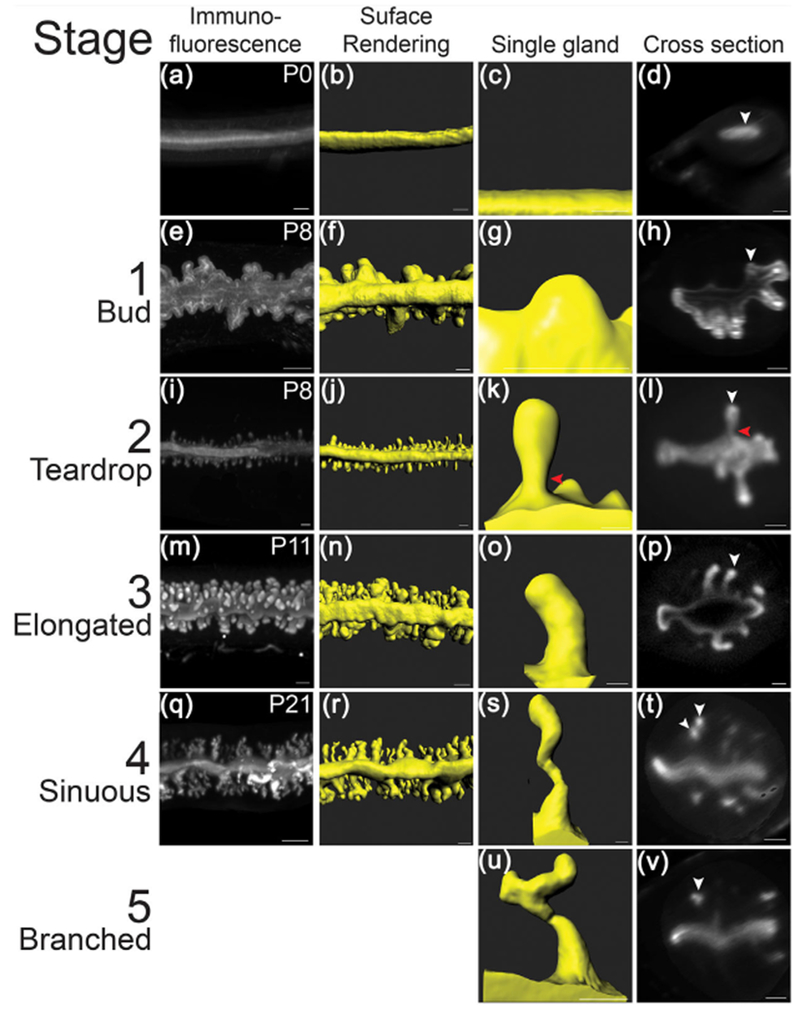
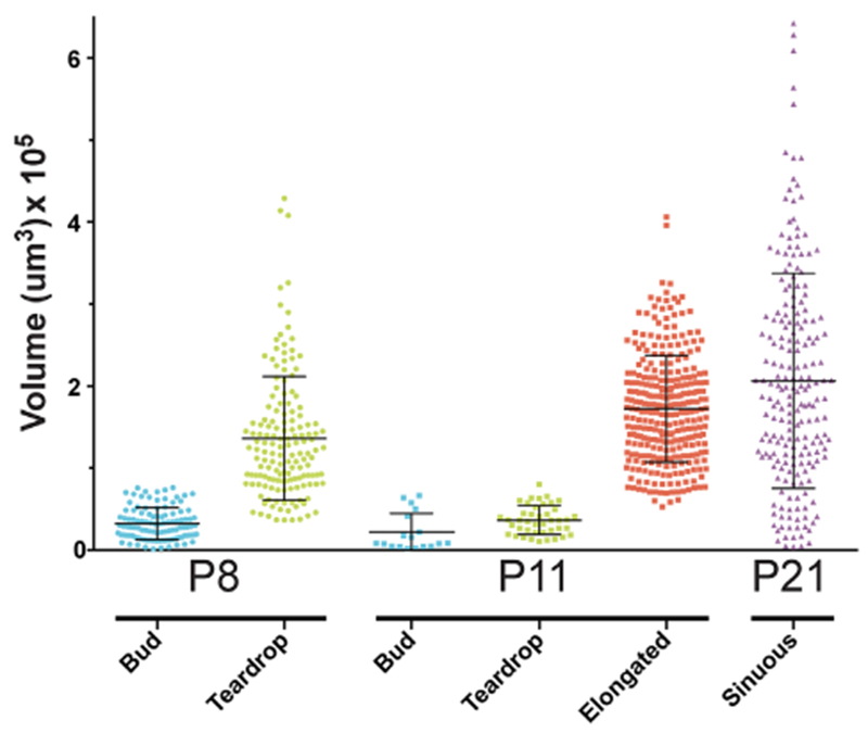
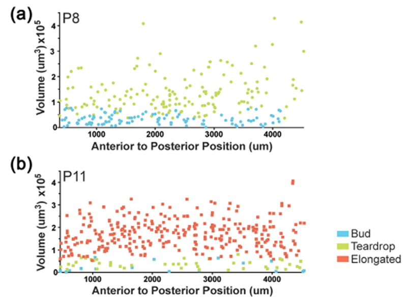
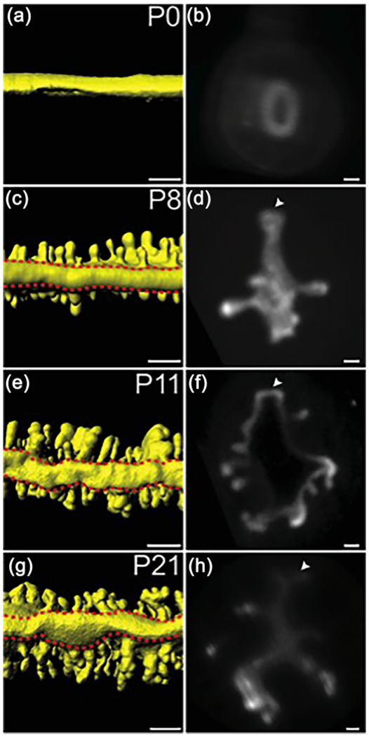
Endometrial or uterine glands secrete substances essential for uterine receptivity to the embryo, implantation, conceptus survival, and growth. Adenogenesis is the process of gland formation within the stroma of the uterus. In the mouse, uterine gland formation initiates at postnatal day (P) 5. Uterine gland morphology is poorly understood because it is primarily based on two-dimensional (2D) histology. To more fully describe uterine gland morphogenesis, we generated three-dimensional (3D) models of postnatal uterine glands from P0 to P21, based on volumetric imaging using light sheet microscopy. At birth (P0), there were no glands. At P8, we found bud- and teardrop-shaped epithelial invaginations. By P11, the forming glands were elongated epithelial tubes. By P21, the elongated tubes had a sinuous morphology. These morphologies are homogeneously distributed along the anterior-posterior axis of the uterus. To facilitate uterine gland analyses, we propose a novel 3D staging system of uterine gland morphology during development in the prepubertal mouse. We define five uterine gland stages: Stage 1: bud; Stage 2: teardrop; Stage 3: elongated; Stage 4: sinuous; and Stage 5: primary branches. This staging system provides a standardized key to assess and quantify prepubertal uterine gland morphology that can be used for studies of uterine gland development and pathology. In addition, our studies suggest that gland formation initiation occurs during P8 and P11. However, between P11 and P21 gland formation initiation stops and all glands elongate and become sinuous. We also found that the mesometrial epithelium develops a unique morphology we term the uterine rail.
Keywords: adenogenesis; epithelium; postnatal development; reproductive tract; uterus.
© 2018 Wiley Periodicals, Inc.
Figures

Similar articles
-
Epithelial morphogenesis in the perinatal mouse uterus.Dev Dyn. 2020 Nov;249(11):1377-1386. doi: 10.1002/dvdy.234. Epub 2020 Sep 3. Dev Dyn. 2020. PMID: 32767478 Free PMC article.
-
Developmental biology of uterine glands.Biol Reprod. 2001 Nov;65(5):1311-23. doi: 10.1095/biolreprod65.5.1311. Biol Reprod. 2001. PMID: 11673245 Review.
-
Ovarian regulation of endometrial gland morphogenesis and activin-follistatin system in the neonatal ovine uterus.Biol Reprod. 2003 Sep;69(3):851-60. doi: 10.1095/biolreprod.103.016337. Epub 2003 May 14. Biol Reprod. 2003. PMID: 12748121
-
Differential Wnt signaling activity limits epithelial gland development to the anti-mesometrial side of the mouse uterus.Dev Biol. 2017 Mar 15;423(2):138-151. doi: 10.1016/j.ydbio.2017.01.015. Epub 2017 Jan 30. Dev Biol. 2017. PMID: 28153546
-
Uterine and placental factors regulating conceptus growth in domestic animals.J Anim Sci. 2004;82 E-Suppl:E4-13. doi: 10.2527/2004.8213_supplE4x. J Anim Sci. 2004. PMID: 15471813 Review.
Cited by
-
Distinguish Characters of Luminal and Glandular Epithelium from Mouse Uterus Using a Novel Enzyme-Based Separation Method.Reprod Sci. 2023 Jun;30(6):1867-1877. doi: 10.1007/s43032-022-01154-z. Epub 2022 Dec 29. Reprod Sci. 2023. PMID: 36581776
-
Single-cell insights into epithelial morphogenesis in the neonatal mouse uterus.Proc Natl Acad Sci U S A. 2023 Dec 5;120(49):e2316410120. doi: 10.1073/pnas.2316410120. Epub 2023 Nov 29. Proc Natl Acad Sci U S A. 2023. PMID: 38019863 Free PMC article.
-
A re-appraisal of mesenchymal-epithelial transition (MET) in endometrial epithelial remodeling.Cell Tissue Res. 2023 Feb;391(2):393-408. doi: 10.1007/s00441-022-03711-z. Epub 2022 Nov 19. Cell Tissue Res. 2023. PMID: 36401092 Free PMC article.
-
Biophysics in oviduct: Planar cell polarity, cilia, epithelial fold and tube morphogenesis, egg dynamics.Biophys Physicobiol. 2019 Feb 26;16:89-107. doi: 10.2142/biophysico.16.0_89. eCollection 2019. Biophys Physicobiol. 2019. PMID: 30923666 Free PMC article. Review.
-
Three-dimensional understanding of the morphological complexity of the human uterine endometrium.iScience. 2021 Mar 2;24(4):102258. doi: 10.1016/j.isci.2021.102258. eCollection 2021 Apr 23. iScience. 2021. PMID: 33796844 Free PMC article.
References
-
- Brody JR, & Cunha GR (1989). Histologic, morphometric, and immunocytochemical analysis of myometrial development in rats and mice: I. Normal development. Am J Anat, 186, 1–20. - PubMed
Publication types
MeSH terms
Grants and funding
LinkOut - more resources
Full Text Sources
Other Literature Sources
