. 2018 Aug 1;315(2):L212-L226.
doi: 10.1152/ajplung.00044.2018. Epub 2018 Mar 15.

Calcitonin gene-related peptide hyperpolarizes mouse pulmonary artery endothelial tubes through KATP channel activation

Affiliations

Calcitonin gene-related peptide hyperpolarizes mouse pulmonary artery endothelial tubes through KATP channel activation

Charles E Norton et al. Am J Physiol Lung Cell Mol Physiol. .

Abstract

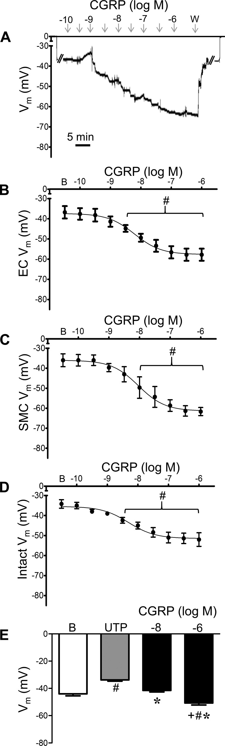
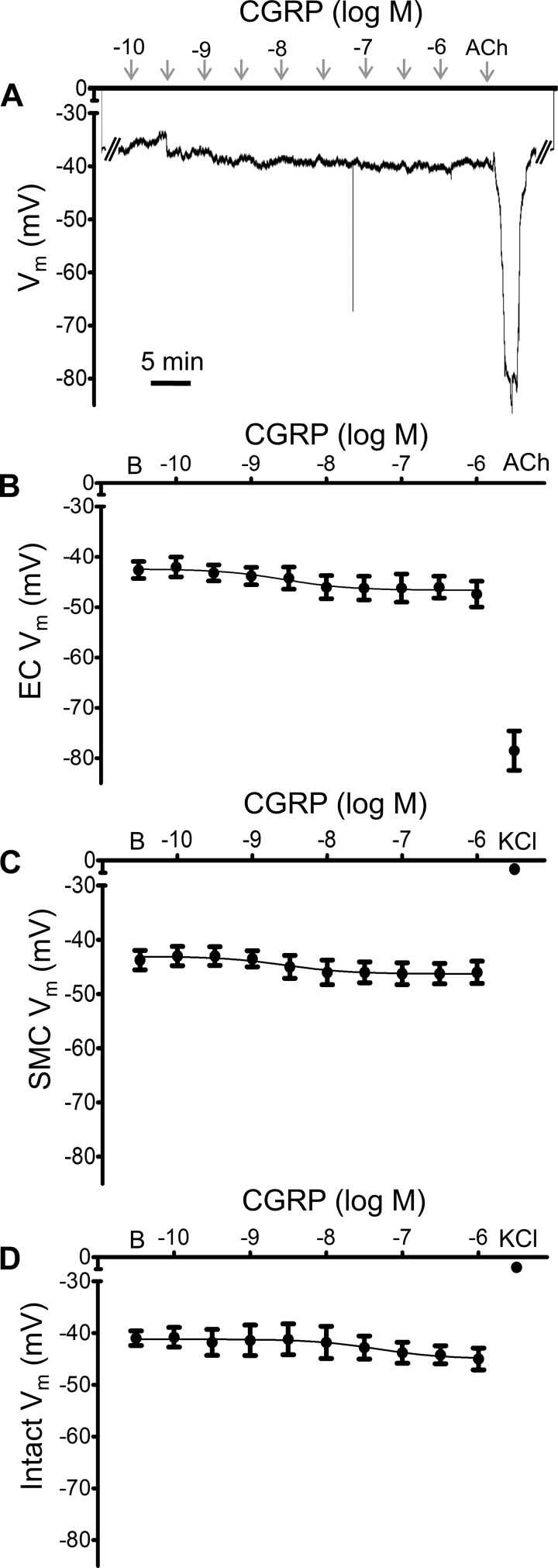
The sensory neurotransmitter calcitonin gene-related peptide (CGRP) is associated with vasodilation of systemic arteries through activation of ATP-sensitive K+ (KATP) channels in smooth muscle cells (SMCs); however, its effects on endothelial cell (EC) membrane potential ( Vm) are unresolved. In pulmonary arteries (PAs) of C57BL/6J mice, we questioned whether CGRP would hyperpolarize ECs as well as SMCs. Intact PAs were isolated and immunostained for CGRP to confirm sensory innervation; vessel segments (1-2 mm long, ∼150 µm diameter) with intact or denuded endothelium were cannulated and pressurized to 16 cmH2O at 37°C. Increasing concentrations (10-10-10-6 M) of CGRP progressively dilated PAs preconstricted with UTP (10-5 M); SMCs hyperpolarized similarly (Δ Vm ∼20 mV) before and after endothelial denudation. To study native intact PA ECs, SMCs were dissociated to isolate endothelial tubes, and their integrity was confirmed by vital dye uptake, nuclear staining, and reproducible electrical and intracellular Ca2+ responses to acetylcholine (10-5 M) over 2 h. Increasing [CGRP] hyperpolarized ECs in a manner similar to SMCs, with each cell layer demonstrating robust immunostaining for CGRP receptor proteins. Increasing concentrations (10-10-10-6 M) of pinacidil, a KATP channel agonist, resulted in progressive hyperpolarization of SMCs of intact PAs (Δ Vm ∼30 mV), which was blocked by glibenclamide (10-6 M), as was hyperpolarization of ECs and SMCs to CGRP. Inhibition of protein kinase A with protein kinase inhibitor (10-5 M) also inhibited hyperpolarization to CGRP. We demonstrate [CGRP]-dependent hyperpolarization of ECs for the first time while validating freshly isolated PA endothelial tubes as an experimental model. Redundant electrical signaling to CGRP in ECs and SMCs implies an integral role for KATP channels in PA dilation.

Keywords: hyperpolarization; lung blood flow; protein kinase A; sensory nerves; vasodilation.


Figures

Fig. 1.
Fig. 1.
Mouse pulmonary arteries are innervated by sensory nerves. Representative calcitonin-gene-related peptide (CGRP) staining (maximum z projections) confirms sensory innervation in larger (~300 μm diameter, A) and smaller (~150 μm diameter, B) pulmonary arteries. Arrows indicate nerve fibers; dotted lines in B indicate vessel edge. Scale bars = 20 µm.
Fig. 2.
Fig. 2.
Calcitonin-gene-related peptide (CGRP) dilates pulmonary arteries (PAs) preconstricted with UTP. A: concentration response for vasoconstriction to UTP (10−9–10−3 M) in isolated, pressurized (16 cmH2O), intact PAs. B: UTP (5 × 10⁻⁶ M) constricted intact PAs, and this effect was not different for endothelium-denuded PAs. C: vasodilation of intact PAs preconstricted with UTP (5 × 10⁻⁶ M) to maximal [ACh] (10−5 M) was abolished following endothelial denudation. D: concentration response for vasodilation to CGRP (10−10–10−6 M) of intact PAs preconstricted with UTP (5 × 10⁻⁶ M). Values are means ± SE; n = 4–5 vessels per group. *P < 0.05 vs. intact. Details: UTP constricted arteries from 126 ± 12 µm inner diameter (ID) at rest to 19 ± 3 µm ID (maximal response). To preconstrict arteries for dilator studies, 5 × 10⁻⁶ M (EC50) UTP constricted intact PAs from 128 ± 8 to 68 ± 6 µm ID. In the presence of UTP, CGRP dilated intact PAs to maximal ID of 119 ± 7 µm.
Fig. 3.
Fig. 3.
Live/dead assay and cell morphology of pulmonary artery (PA) endothelial tubes. A: calcein staining of cytoplasm of live cells. B: ethidium bromide staining of dead cells beneath pinning pipettes (located at each end of the endothelial tube in respective panels). C: overlay image of A and B. D: bright-field image of boxed region in C shows endothelial cell (EC) orientation and intact morphology of endothelial tube secured between pinning pipettes. E: staining with Hoechst 33342 illustrates smooth muscle cell nuclei (thin, vertical) and EC nuclei (oval, horizontal) of an intact pressurized PA. F: nuclei staining showing purity for EC composition of endothelial tube. Apparent increase in EC nuclei compared with E reflects “flattened” tube with top and bottom sides within the same focal plane. Scale bars = 50 µm.
Fig. 4.
Fig. 4.
Stability of electrical and Ca2+ signaling in pulmonary artery (PA) endothelial tubes. A: representative continuous recording for membrane potential (Vm) and intracellular Ca2+ concentration ([Ca2+]i) responses (see methods) to intermittent exposure to ACh (10−5 M) during a 2-h protocol. Horizontal bars indicate periods of ACh superfusion. B: summary data. Pre, before ACh; ACh, peak hyperpolarization; Post, at 5-min washout. Values are means ± SE; n = 5 per group.
Fig. 5.
Fig. 5.
Pulmonary artery (PA) smooth muscle cells (SMCs) and endothelial cells (ECs) are hyperpolarized by calcitonin gene-related peptide (CGRP). A: representative continuous recording of membrane potential (Vm) illustrates concentration-dependent hyperpolarization of SMCs by CGRP in a denuded PA. From baseline (zero), note abrupt change in Vm upon entry and exit from cell. W, washout. B: summary data for Vm of PA endothelial tubes to CGRP. B, baseline. C: summary data for Vm of SMCs of denuded PAs to CGRP. D: summary data for Vm of SMCs of intact PAs to CGRP. E: summary data for Vm during resting baseline, with 5 × 10⁻⁶ M UTP alone and during addition of 10−8 or 10−6 M CGRP. Values are means ± SE; n = 5 per group. #P < 0.05 vs. baseline Vm. *P < 0.05 vs. UTP. +P < 0.05 vs. 10−8 M CGRP. UTP depolarized PAs 10 ± 1 mV, from which 10−6 M CGRP hyperpolarized PAs 17 ± 2 mV.
Fig. 6.
Fig. 6.
Immunostaining for calcitonin receptor proteins in pulmonary arteries (PAs). Representative dual staining of PAs for receptor activity-modifying protein 1 (RAMP1, left, green) and calcitonin receptor-like receptor (CRLR; middle, red) confirms CRLR expression in endothelial cells (ECs) and smooth muscle cells (SMCs). Overlay images (right) illustrate corresponding localization (yellow) of respective proteins. Scale bar = 20 µm.
Fig. 7.
Fig. 7.
ATP-sensitive K+ channel activation hyperpolarizes pulmonary artery (PA) endothelial cells (ECs) and smooth muscle cells (SMCs). A: representative continuous recording of membrane potential (Vm) illustrates concentration-dependent hyperpolarization of ECs to pinacidil (10−9−10−5 M) in a PA endothelial tube. W, washout. B: summary data for hyperpolarization to pinacidil in endothelial tubes. C: summary data for hyperpolarization to pinacidil in SMCs of intact PAs. Values are means ± SE; n = 4 per group. B, baseline Vm. #P < 0.05 vs. baseline Vm. EC50 values for pinacidil were 7 × 10⁻⁸ M in ECs and 4 × 10⁻⁸ M in SMCs.
Fig. 8.
Fig. 8.
Glibenclamide reverses ATP-sensitive K+ channel-dependent hyperpolarization of pulmonary artery (PA) endothelial cells (ECs) and smooth muscle cells (SMCs). A: representative continuous recording of membrane potential (Vm) from an endothelial tube illustrates concentration-dependent reversal of hyperpolarization to pinacidil (P; 10−5 M, maintained throughout) by glibenclamide. B: summary data for reversal of hyperpolarization to pinacidil in endothelial tubes with glibenclamide (10−10−10−6 M). C: summary data for reversal of hyperpolarization to pinacidil in SMCs of intact PAs with glibenclamide (10−10−10−6 M). Values are means ± SE; n = 4 per group. B, baseline Vm; P, Vm during 10−5 M pinacidil. #P < 0.05 vs. baseline Vm. EC50 values for glibenclamide were 6 × 10⁻⁹ M in ECs and 2 × 10⁻⁸ M in SMCs.
Fig. 9.
Fig. 9.
Hyperpolarization to calcitonin gene-related peptide (CGRP) in pulmonary artery (PA) endothelial cells (ECs) and smooth muscle cells (SMCs) requires ATP-sensitive K+ (KATP) channels. A: representative continuous recording of membrane potential (Vm) from an endothelial tube in the presence of glibenclamide (10−6 M) illustrates lack of responses to CGRP (10−10−10−6 M) with maintenance of hyperpolarization to ACh (10−5 M; 5 min). B: summary data for loss of hyperpolarization to CGRP (but not to ACh) for endothelial tubes in the presence of glibenclamide (10−6 M). C: summary data for loss of hyperpolarization to CGRP for SMCs of denuded PAs; note final depolarization with KCl (10−1 M). D: summary data for loss of hyperpolarization to CGRP (but not depolarization to KCl) for SMCs of intact PAs. Values are means ± SE; n = 4–5 per group. B, baseline Vm. Details: inhibition of KATP channels also attenuated hyperpolarization in female mice: ΔVm = 3 ± 1 mV for ECs and 4 ± 2 mV for SMCs from intact PAs (n = 3 per group). To confirm electrode placement, from a resting Vm of −43 ± 2 mV, ACh hyperpolarized ECs to −79 ± 4 mV (n = 4), while KCl depolarized SMCs from a resting Vm of −41 ± 3 to −9 ± 3 mV in intact PAs (n = 4) and to −7 ± 2 mV in denuded PAs (n = 2).
Fig. 10.
Fig. 10.
Genetic deletion of ATP-sensitive K+ (KATP) channels inhibits hyperpolarization to calcitonin gene-related peptide (CGRP). A: representative continuous recording of membrane potential (Vm) from endothelial tube of a mouse lacking KATP channels illustrates absence of responses to CGRP (10−10−10−6 M) or pinacidil (P; 10−5 M) followed by hyperpolarization to ACh (10−5 M; 5 min). B: summary data for responses to CGRP in smooth muscle cells of intact pulmonary arteries from KATP−/− mice. Values are means ± SE; n = 3 per group. B, baseline Vm. #P < 0.05 vs. baseline Vm.
Fig. 11.
Fig. 11.
Calcitonin gene-related peptide (CGRP)-dependent hyperpolarization in endothelial cells (ECs) and smooth muscle cells (SMCs) requires PKA. A: representative continuous recording of membrane potential (Vm) from a SMC of an intact pulmonary artery (PA) in the presence of PKI (10−5 M) illustrates lack of response to CGRP (10−10−10−6 M) followed by depolarization to KCl (10−1 M; 5 min). B: summary data for Vm during exposure of endothelial tubes to CGRP in the presence of PKI (10−5 M) followed by hyperpolarization to ACh (10−5 M). C: summary data for Vm during exposure of SMCs of denuded PAs to CGRP followed by depolarization to KCl (10−1 M). D: summary data for Vm during exposure of intact PAs to CGRP followed by depolarization to KCl (10−1 M). ACh hyperpolarized ECs by ∼36 mV, and KCl depolarized SMCs by ∼30 mV. Values are means ± SE; n = 4 per group. B,  baseline Vm. #P < 0.05 vs. baseline Vm.
Fig. 12.
Fig. 12.
Roles for ATP-sensitive K+ channels and PKA in pulmonary artery (PA) dilation to calcitonin gene-related peptide (CGRP). A: vasodilation during EC50 (10−8 M) and maximal (10−6 M) [CGRP] for intact PAs preconstricted with UTP (5 × 10⁻⁶ M) in the absence (vehicle) and presence of glibenclamide (10−6 M). B: changes in vessel wall [Ca2+]i (ΔF340/F380; see methods) of intact PAs corresponding with A. C: vasodilation to CGRP in the absence and presence of PKI (10−5 M). D: changes in vessel wall [Ca2+]i (ΔF340/F380) corresponding with C. Values are means ± SE; n = 4–5 per group. #P < 0.05 vs. vehicle. +P < 0.05 vs. 10−8 M CGRP. Details: in A and B, baseline diameters [137 ± 9 and 134 ± 8 µm in the absence (vehicle) and presence of glibenclamide, respectively] and resting [Ca2+]i (F340/F380 = 0.51 ± 0.05 and 0.57 ± 0.06, respectively) were similar between groups. Responses to UTP were not significantly different between PAs during superfusion with respective treatments [inner diameter (ID) = 72 ± 5 µm, constriction = 46 ± 4%, and ΔF340/F380 = 0.24 ± 0.05 for vehicle; ID = 71 ± 10 µm, constriction = 47 ± 6%, and ΔF340/F380 = 0.21 ± 0.04 for glibenclamide]. C and D: baseline diameters (134 ± 4 and 124 ± 11 µm for vehicle and PKI, respectively), Ca2+ (F340/F380 = 0.63 ± 0.06 and 0.60 ± 0.08 for vehicle and PKI, respectively), and responses to UTP (ID = 75 ± 5 µm, constriction = 43 ± 3%, and ΔF340/F380 = 0.17 ± 0.02 for vehicle; ID = 64 ± 8 µm, constriction = 47 ± 2%, and ΔF340/F380 = 0.20 ± 0.06 for PKI) were similar between conditions.
Fig. 13.
Fig. 13.
Pulmonary artery (PA) dilation to calcitonin gene-related peptide (CGRP) has both endothelial and smooth muscle components. A: vasodilation to CGRP of UTP-preconstricted (5 × 10⁻⁶ M) intact (±l-NAME, 10−4 M) and endothelium-denuded PAs during EC50 (10−8 M) and maximal (10−6 M) [CGRP]. B: changes in vessel wall [Ca2+]i (ΔF340/F380; see methods) in response to CGRP were similar for intact (±l-NAME) and denuded arteries. Values are means ± SE; n = 4 per group. #P < 0.05 vs. intact. +P < 0.05 vs. 10−8 M CGRP. Details: baseline diameter = 132 ± 11, 133 ± 6, and 128 ± 4 µm for intact, denuded, and l-NAME, respectively; resting intracellular [Ca2+] (F340/F380) = 0.62 ± 0.04, 0.58 ± 0.04, and 0.57 ± 0.02 for intact, denuded, and l-NAME, respectively. Responses to 5 × 10⁻⁶ M UTP (ID = 76 ± 11 µm, constriction = 44 ± 5%, and ΔF340/F380 = 0.19 ± 0.03 (intact); ID = 66 ± 10 µm, constriction = 47 ± 7%, and ΔF340/F380 = 0.20 ± 0.02 (denuded); and ID = 68 ± 3 µm, constriction = 47 ± 3%, and ΔF340/F380 = 0.15 ± 0.04 (l-NAME)] were not significantly different between groups.

Similar articles

Cited by

References

    1. Archer SL, Huang JM, Hampl V, Nelson DP, Shultz PJ, Weir EK. Nitric oxide and cGMP cause vasorelaxation by activation of a charybdotoxin-sensitive K channel by cGMP-dependent protein kinase. Proc Natl Acad Sci USA 91: 7583–7587, 1994. doi:10.1073/pnas.91.16.7583. - DOI - PMC - PubMed
    1. Babenko AP, Aguilar-Bryan L, Bryan J. A view of sur/KIR6.X, KATP channels. Annu Rev Physiol 60: 667–687, 1998. doi:10.1146/annurev.physiol.60.1.667. - DOI - PubMed
    1. Brain SD, Grant AD. Vascular actions of calcitonin gene-related peptide and adrenomedullin. Physiol Rev 84: 903–934, 2004. doi:10.1152/physrev.00037.2003. - DOI - PubMed
    1. Bartosik I, Eskilsson J, Ekman R, Akesson A, Scheja A. Correlation between plasma concentrations of calcitonin gene related peptide and pulmonary pressure in patients with systemic sclerosis. Ann Rheum Dis 61: 261–263, 2002. doi:10.1136/ard.61.3.261. - DOI - PMC - PubMed
    1. Beech DJ, Zhang H, Nakao K, Bolton TB. K channel activation by nucleotide diphosphates and its inhibition by glibenclamide in vascular smooth muscle cells. Br J Pharmacol 110: 573–582, 1993. doi:10.1111/j.1476-5381.1993.tb13849.x. - DOI - PMC - PubMed

Publication types

LinkOut - more resources