. 2018 Jun 1;314(6):G677-G689.
doi: 10.1152/ajpgi.00381.2017. Epub 2018 Mar 15.

Primary cilia disruption differentially affects the infiltrating and resident macrophage compartment in the liver

Affiliations

Primary cilia disruption differentially affects the infiltrating and resident macrophage compartment in the liver

Kurt A Zimmerman et al. Am J Physiol Gastrointest Liver Physiol. .

Abstract

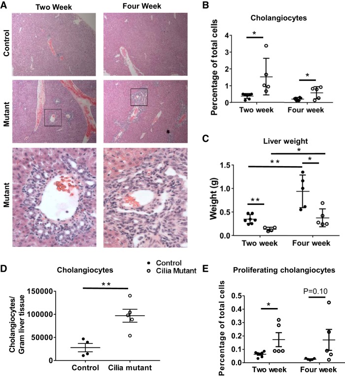
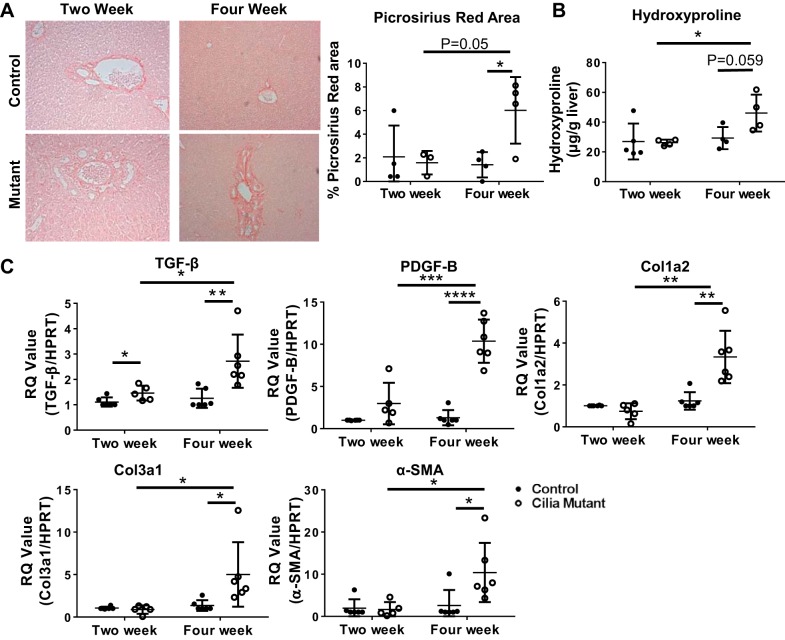
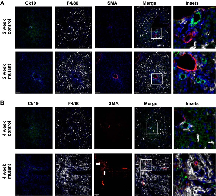
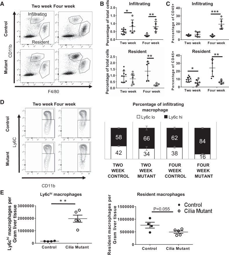
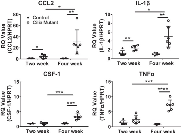
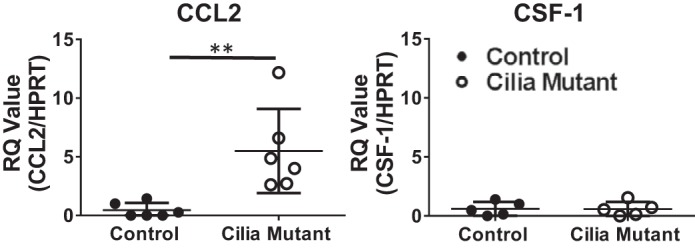
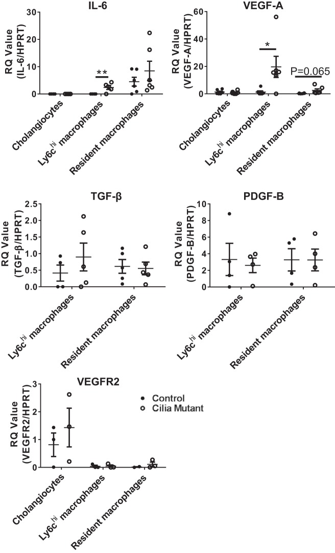
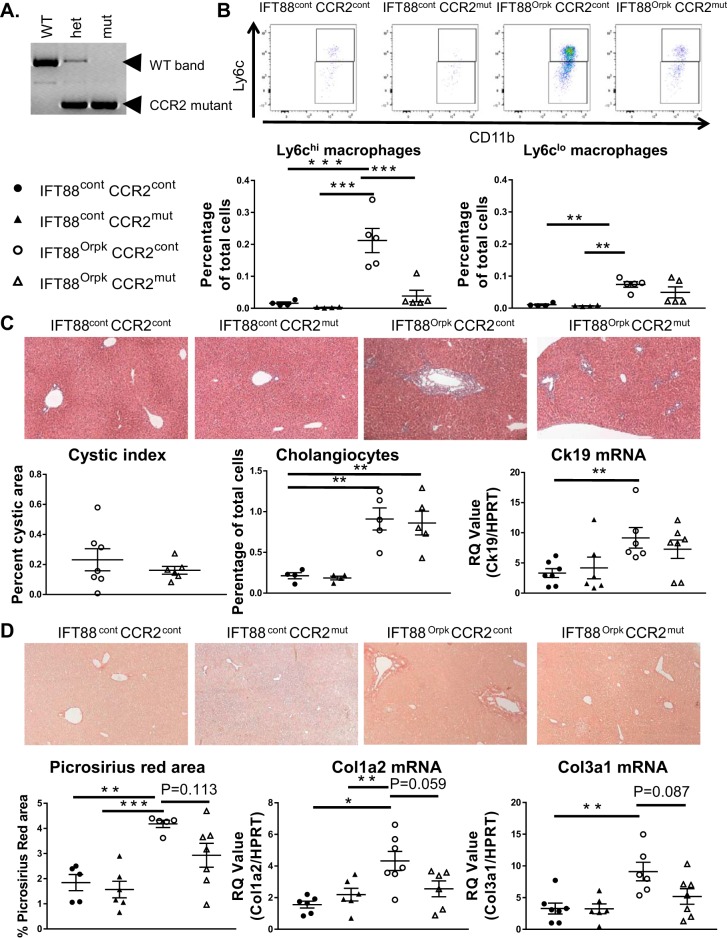
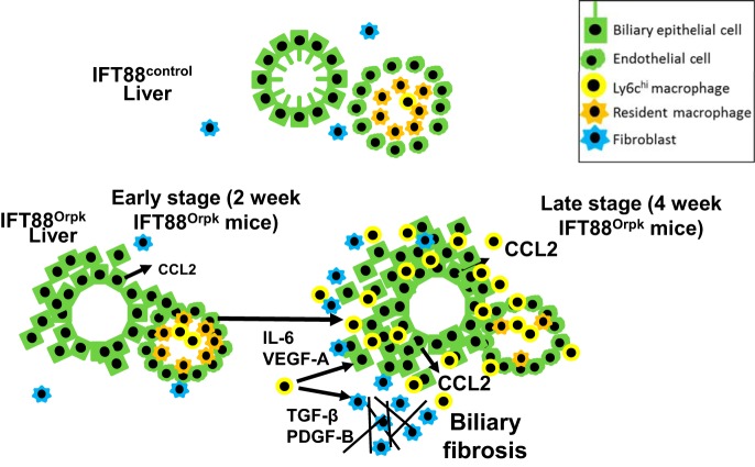
Hepatorenal fibrocystic disease (HRFCD) is characterized by cysts in the kidney and liver with associated fibrosis and is the result of defects in proteins required for cilia function or assembly. Previous reports indicate that macrophages, mainly M2-like macrophages, contribute to HRFCD, although the origin of these cells (yolk sac-derived resident macrophages vs. bone marrow-derived infiltrating macrophages) and their contribution to the observed phenotypes are unknown. We utilize a congenital model of cilia dysfunction (IFT88Orpk) to study the importance of macrophages in HRFCD. Our data show a rapid expansion of the bile duct region and development of fibrosis between 2 and 4 wk of age. Immunofluorescence microscopy analysis reveals an accumulation of F4/80+ macrophages in regions exhibiting biliary hyperplasia in IFT88Orpk mice. Flow cytometry data show that cilia dysfunction leads to an accumulation of infiltrating macrophages (CD11bhi, F4/80lo) and a reduction of resident macrophage (CD11blo, F4/80hi) number. A majority of the infiltrating macrophages are Ly6chi profibrogenic macrophages. Along with the accumulation of immune cells, expression of proinflammatory and profibrotic transcripts, including TGF-β, TNF-α, IL-1β, and chemokine (C-C) motif ligand 2, is increased. Quantitative RT-PCR analysis of flow-sorted cells shows enhanced expression of CCL2 in cholangiocytes and enhanced expression of VEGF-A and IL-6 in Ly6chi macrophages. Genetic inhibition of Ly6chi macrophage accumulation in IFT88Orpk FVB CCR2-/- mice reduced biliary fibrosis but did not affect epithelial expansion. Collectively, these studies suggest that biliary epithelium with defects in primary cilia preferentially recruits Ly6chi infiltrating macrophages, which promote fibrotic progression in HRFCD pathogenesis. NEW & NOTEWORTHY These studies are the first to address the contribution of the infiltrating and resident macrophage niche during progression of hepatorenal fibrocystic disease (HRFCD). We show that the number of infiltrating macrophages is significantly upregulated in HRFCD mouse models. Finally, we show that prevention of Ly6chi infiltrating macrophage accumulation significantly reduces biliary fibrosis, but not biliary hyperplasia, suggesting that this population may be responsible for the fibrotic progression of the disease in HRFCD patients.

Keywords: cholangiocyte; hepatorenal fibrocystic disease; liver; macrophage.


Figures

Fig. 1.
Fig. 1.
Rapid expansion of the biliary region in cilia mutant mice between 2 and 4 wk of age. A: hematoxylin-eosin-stained sections of liver tissue harvested from control and cilia mutant mice at 2 and 4 wk of age. Inset regions within black boxes in top mutant images are shown in bottom mutant images. B: quantification of cholangiocyte number in 2- and 4-wk-old mice (n = 5–10) as determined by flow cytometry for cytokeratin 19-positive cells. C: liver weight of control and cilia mutant livers at 2 and 4 wk (n = 5–7 mice). D: quantification of absolute numbers of cholangiocytes in control and cilia mutant livers at 4 wk (n = 4–5 mice). E: number of proliferating cholangiocytes as a percentage of total cells quantified using flow cytometry staining for Ki67, cytokeratin 19 double-positive cells (n = 5–7 mice). Values are means ± SE. *P < 0.05; **P < 0.01.
Fig. 2.
Fig. 2.
IFT88Orpk mice develop fibrosis in periportal regions at 4 wk of age. A: picrosirius red-stained sections of liver tissue from 2- and 4-wk-old control and cilia mutant mice. B: quantification of picrosirius red-stained area (n = 3–4 mice) and hydroxyproline assay of livers from 2- and 4-wk-old mice (n = 4–5 mice). C: quantitative RT-PCR determination of TGF-β, PDGF-B, collagen types 1 and 3 (Col1a2 and Col3a1), and α-smooth muscle actin (SMA) expression in mRNA isolated from whole liver of 2- and 4-wk-old control and IFT88Orpk mice (n = 5–8). For each gene analyzed, expression levels in control mice were set to 1. RQ, relative quantification; HPRT, hypoxanthine phosphoribosyltransferase. Values are means ± SE. *P < 0.05; **P < 0.01; ***P < 0.001; ****P < 0.0001.
Fig. 3.
Fig. 3.
F4/80+ macrophages accumulate in regions of α-smooth muscle actin (SMA)-positive myofibroblasts and cytokeratin 19 (Ck19)-positive cholangiocytes in 4-wk-old IFT88Orpk mice. Livers were harvested from 2- and 4-wk-old control and IFT88Orpk mice (n = 4–6) and stained with cytokeratin 19 (cholangiocytes), F4/80 (pan-macrophage marker), and SMA (myofibroblasts). A: immunofluorescence confocal microscopy shows mild accumulation of F4/80+ cells in regions of cholangiocyte expansion in 2-wk-old IFT88Orpk mutant mice. Inset region is depicted with a white box. B: immunofluorescence confocal microscopy shows severe accumulation of F4/80+ macrophages (white) in regions of cholangiocyte (green) expansion in IFT88Orpk mice. Accumulation of SMA+ myofibroblasts (red) in regions containing cholangiocyte expansion and macrophages suggests possible communication between these cells. A representative image is shown for each group of mice. Inset region is depicted with a white box. Arrows denote region containing numerous SMA+ myofibroblasts.
Fig. 4.
Fig. 4.
Flow cytometry reveals a shift in macrophage profile in IFT88Orpk mice during postnatal liver development and an accumulation of Ly6chi macrophages in IFT88Orpk mice before cholangiocyte expansion and periportal fibrosis. Livers were harvested from control and IFT88Orpk mice at 2 and 4 wk of age, stained, and live single cells were analyzed based on surface expression of CD11b and F4/80. A: representative fluorescence-activated cell sorting (FACS) plot is shown. B and C: quantification of infiltrating and resident macrophages gated as a percentage of total cells and as a percentage of immune cells (CD45+) (n = 5–10 mice). D: representative FACS plot of infiltrating macrophages that were further subgated based on expression of Ly6c. Ly6chi macrophages were quantified as a percentage of total infiltrating macrophages (n = 5–10). E: whole livers from 4-wk-old control and IFT88Orpk mice (n = 4–6) were sorted, and absolute numbers of Ly6chi infiltrating macrophages and tissue resident macrophages were determined by flow cytometry. Cell numbers were normalized to average liver weight, as shown in Fig. 1C. Values are means ± SE. *P < 0.05; **P < 0.01; ***P < 0.001.
Fig. 5.
Fig. 5.
Expression of proinflammatory and profibrotic cytokines is increased in IFT88Orpk mice. Livers from 2- and 4-wk-old mice were harvested and minced, and mRNA was extracted using TRIzol. Transcript levels of chemokine (C-C) motif ligand 2 (CCL2), IL-1β, colony-stimulating factor 1 (CSF-1), and TNF-α from whole liver tissue were determined by quantitative RT-PCR and normalized to the housekeeping gene hypoxanthine phosphoribosyltransferase (HPRT). For each gene, expression levels in 2-wk-old control mice were set to 1. RQ, relative quantification. Values are means ± SE; n = 5–7. *P < 0.05; **P < 0.01; ***P < 0.001; ****P < 0.0001.
Fig. 6.
Fig. 6.
Expression of chemokine (C-C) motif ligand 2 (CCL2) is increased in epithelial cells from IFT88Orpk mice. Expression of CCL2 and colony-stimulating factor 1 (CSF-1) in fluorescence-activated cell-sorted control and cilia mutant epithelial cells was determined by quantitative RT-PCR. RQ, relative quantification; HPRT, hypoxanthine phosphoribosyltransferase. Values are means ± SE; n = 6. **P < 0.01.
Fig. 7.
Fig. 7.
Increased gene expression of proinflammatory and profibrotic cytokines in Ly6chi macrophages from IFT88Orpk mice. Gene expression of proproliferative cytokines (IL-6 and VEGF-A), profibrotic growth factors (TGF-β and PDGF-B), and Kdr, which encodes VEGF receptor 2 (VEGFR2), in cholangiocytes, infiltrating macrophages, and resident macrophages isolated from the liver of 4-wk-old control and cilia mutant mice was determined by quantitative RT-PCR. RQ, relative quantification; HPRT, hypoxanthine phosphoribosyltransferase. Values are means ± SE; n = 5–6. *P < 0.05; **P < 0.01.
Fig. 8.
Fig. 8.
Ly6chi macrophages and fibrosis are reduced in IFT88Orpk FVB CCR2−/− mice. A: PCR analysis of DNA extracted from wild-type (WT), heterozygous (het), and knockout (mut) mice for the CCR2 gene. B: representative fluorescence-activated cell sorting plots of infiltrating macrophage subtypes harvested from control (cont) and IFT88Orpk (mut) mice on control (cont) or CCR−/− (mut) background at 4 wk of age with quantification of Ly6chi and Ly6clo macrophages from their respective groups (n = 4–5). C: representative histological images of liver sections from 4-wk-old mice (n = 6–8). Cystic index was quantified for IFT88Orpk CCR2 control and IFT88Orpk CCR2−/− mice. Cholangiocyte number was quantified using flow cytometry [(cytokeratin 19 (Ck19)-positive cells] and quantitative RT-PCR for Ck19. D: representative picrosirius red-stained sections from 4-wk-old mice (n = 6–8) with quantification of picrosirius red-positive area and quantitative RT-PCR data for collagen types 1 and 3 (Col1a2 and Col3a1). Values are means ± SE. *P < 0.05; **P < 0.01; ***P < 0.001.
Fig. 9.
Fig. 9.
Proposed mechanism responsible for enhanced bile duct expansion and fibrosis in IFT88Orpk liver. Schematic depicts reciprocal signaling between bile duct cholangiocytes, macrophages, and myofibroblasts during development of hepatic fibrocystic liver disease. In our model, the presence of dysfunctional primary cilia on cholangiocytes leads to persistent low levels of injury, increased production of macrophage chemoattractant cytokines [chemokine (C-C) motif ligand 2 (CCL2)], and increased numbers of Ly6chi infiltrating macrophages. Increased numbers Ly6chi infiltrating macrophages promote expansion of biliary fibrosis in 4-wk-old IFT88Orpk mice.

References

    1. Amura CR, Brodsky KS, Groff R, Gattone VH, Voelkel NF, Doctor RB. VEGF receptor inhibition blocks liver cyst growth in pkd2WS25/− mice. Am J Physiol Cell Physiol 293: C419–C428, 2007. doi:10.1152/ajpcell.00038.2007. - DOI - PubMed
    1. Behrens A, Sibilia M, David JP, Möhle-Steinlein U, Tronche F, Schütz G, Wagner EF. Impaired postnatal hepatocyte proliferation and liver regeneration in mice lacking c-jun in the liver. EMBO J 21: 1782–1790, 2002. doi:10.1093/emboj/21.7.1782. - DOI - PMC - PubMed
    1. Brempelis KJ, Crispe IN. Infiltrating monocytes in liver injury and repair. Clin Transl Immunology 5: e113, 2016. doi:10.1038/cti.2016.62. - DOI - PMC - PubMed
    1. Chen LP, Qian YY, Li ZL, Bai HW, Cai M, Shi BY. [Role of IL-6/STAT3 in rat cholangiocyte proliferation induced by lipopolysaccharide]. Zhonghua Gan Zang Bing Za Zhi 17: 374–377, 2009. - PubMed
    1. Cowley BD Jr, Gudapaty S, Kraybill AL, Barash BD, Harding MA, Calvet JP, Gattone VH II. Autosomal-dominant polycystic kidney disease in the rat. Kidney Int 43: 522–534, 1993. doi:10.1038/ki.1993.79. - DOI - PubMed

Publication types

MeSH terms