

Case Reports
doi: 10.1371/journal.pntd.0006171.
eCollection 2018 Mar.
The diagnosis and treatment introspection of the first imported case of atypical cerebral schistosomiasis in Guangzhou city
Affiliations
- PMID: 29543824
- PMCID: PMC5854235
- DOI: 10.1371/journal.pntd.0006171
Item in Clipboard
Case Reports
The diagnosis and treatment introspection of the first imported case of atypical cerebral schistosomiasis in Guangzhou city
PLoS Negl Trop Dis.
.
No abstract available
Conflict of interest statement
The authors have declared that no competing interests exist.
Figures
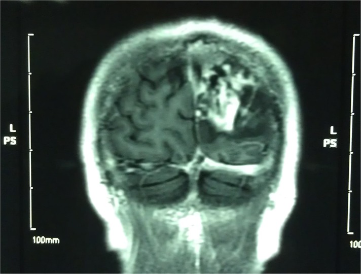
MRI of the brain before treatment shows an occupied lesion in the left occipital region.
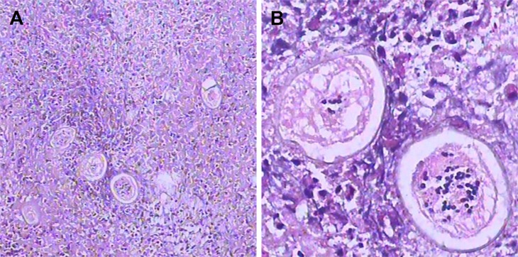
(A) Low-magnification view of the paraffin portion of the formalin-fixed temporal lobe biopsy shows an immature Schistosoma egg surrounded by granuloma and glial cells. (B) High magnification shows two eggs with shells displaying the diagnostic acentric spine shape of immature Schistosoma eggs. Histiocytes near the eggs highlight that the nuclei of Schistosoma are smaller than those of the host’s cells.
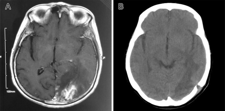
(A) MRI before surgery showing an intracranial occupying lesion in the left occipital lobe, with edema and mottled nodular linear enhancement in the surrounding region. (B) CT result three months after surgery showing the disappearance of the nidus and fading of the edema. CT, computed tomography.
References
-
- CDC. Neglected Tropical Diseases 2011 [cited 2017 July 24]. Available from: https://www.cdc.gov/globalhealth/ntd/diseases/schisto_burden.html.
-
- Colley DG, Bustinduy AL, Secor WE, King CH. Human schistosomiasis. Lancet. 2014;383(9936):2253–64. doi: 10.1016/S0140-6736(13)61949-2 - DOI - PMC - PubMed
-
- Neill PJ, Smith JH, Doughty BL, Kemp M. The ultrastructure of the Schistosoma mansoni egg. Am J Trop Med Hyg. 1988;39(1):52–65. - PubMed
-
- Vale T C, de Sousa-Pereira S R, Ribas J O G R.Lambertucci J R.Neuroschistosomiasismansoni: Literature Review and Guidelines.2012;18:333–342. - PubMed
Publication types
MeSH terms
Substances
LinkOut - more resources
Full Text Sources
Other Literature Sources
Medical
