

Quality control of malaria microscopy reveals misdiagnosed non-falciparum species and other microscopically detectable pathogens in Senegal
- PMID: 29544479
- PMCID: PMC5853095
- DOI: 10.1186/s12941-018-0261-1
Quality control of malaria microscopy reveals misdiagnosed non-falciparum species and other microscopically detectable pathogens in Senegal
Abstract
Background: In developing countries, malaria diagnosis relies on microscopy and rapid diagnostic tests. In Senegal, national malaria control program (NMCP) regularly conducts supervisory visits in health services where malaria microscopy is performed. In this study, expert microscopists assessed the performance of laboratory technicians in malaria microscopy.
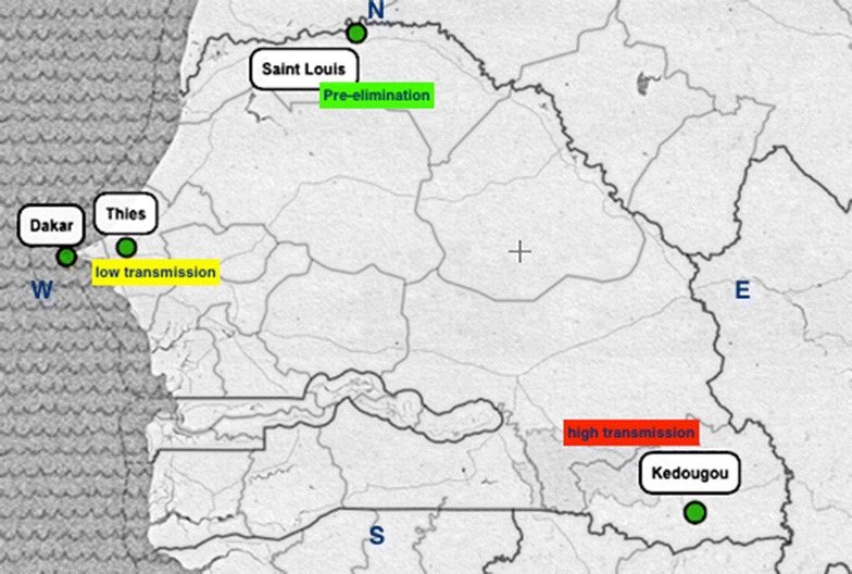
Methods: The present external quality assessment (EQA) was conducted in three different areas of malaria transmission. Participants were laboratory technicians previously trained by NMCP on malaria microscopy. Stored read slides were randomly collected for blinded re-checking by expert microscopists. At the same time a set of 8 slides (3 positive P. falciparum and 5 negative slides) were submitted to participants for proficiency testing. Microscopists performance were evaluated on the basis of the errors rates on slide reading-high false positive (HFP), high false negative (HFN), low false positive (LFP) and low false negative (LFN)-and the calculation of their sensitivities and specificities relative to expert microscopy. Data were entered and analysed using Microsoft Excel software.
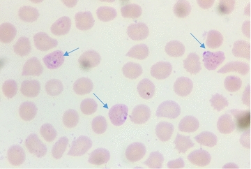
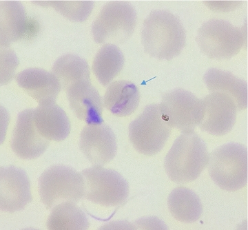
Results: A total of 450 stored slides were collected from 17 laboratories for re-checking. Eight laboratories scored 100% of correct reading. Only one major error was recorded (HFP). Six laboratories recorded LFN results: Borrelia, P. ovale, and low parasite densities (95 and 155 p/μl) were missed. Two P. falciparum slides were misidentified as P. malariae and one P. ovale slide as P. vivax. The overall sensitivities and specificities for all participants against expert microscopists were 97.8 and 98.2% respectively; Sensitivities and specificities of hospital microscopists (96.7 and 98.9%) were statistically similar to those of health centre microscopists (98.5 and 97.8% respectively) (p = 0.3993 and p = 0.9412 respectively). Overall, a very good agreement was noted with kappa value of 0.96 (CI95% 93.4-98.6%) relative to expert microscopy. Proficiency testing showed that among the 17 participants, 11 laboratories scored 100% of correct reading. Three LFN and four LFP results were recorded respectively. The P. falciparum slide with Maurer dots was misidentified as P. ovale in 1 centre and the same slide was misread as P. vivax in another centre; No major error (HFP or HFN) was noted.
Conclusion: EQA of malaria microscopy showed an overall good performance especially regarding P. falciparum detection. However, efforts need to be made addressing the ability to detect non-falciparum species and others endemic blood pathogens such as Borrelia. The further NMCP training sessions and evaluations should consider those aspects to expect high quality-assured capacity for malaria microscopy.
Keywords: Diagnosis; EQA; External quality assessment; Malaria; Microscopy; Senegal.
Figures
Similar articles
-
Performance of malaria microscopy external quality assessment and networking among health facilities in west Amhara region, Ethiopia.BMC Infect Dis. 2020 May 19;20(1):355. doi: 10.1186/s12879-020-05077-5. BMC Infect Dis. 2020. PMID: 32429860 Free PMC article.
-
Performance evaluation of laboratory professionals on malaria microscopy in Hawassa Town, Southern Ethiopia.BMC Res Notes. 2014 Nov 25;7:839. doi: 10.1186/1756-0500-7-839. BMC Res Notes. 2014. PMID: 25422030 Free PMC article.
-
Factors associated with malaria microscopy diagnostic performance following a pilot quality-assurance programme in health facilities in malaria low-transmission areas of Kenya, 2014.Malar J. 2017 Sep 13;16(1):371. doi: 10.1186/s12936-017-2018-2. Malar J. 2017. PMID: 28903758 Free PMC article.
-
Malaria rapid diagnostic tests in endemic settings.Clin Microbiol Infect. 2013 May;19(5):399-407. doi: 10.1111/1469-0691.12151. Epub 2013 Feb 25. Clin Microbiol Infect. 2013. PMID: 23438048 Review.
-
Malaria rapid diagnostic tests in travel medicine.Clin Microbiol Infect. 2013 May;19(5):408-15. doi: 10.1111/1469-0691.12152. Epub 2013 Feb 1. Clin Microbiol Infect. 2013. PMID: 23373854 Review.
Cited by
-
Differential ex vivo susceptibility of Plasmodium malariae and Plasmodium falciparum clinical isolates from Ghana and Mali to current and lead discovery candidate antimalarial drugs.Microbiol Spectr. 2025 Apr;13(4):e0217624. doi: 10.1128/spectrum.02176-24. Epub 2025 Mar 10. Microbiol Spectr. 2025. PMID: 40062863 Free PMC article.
-
Significance of nested PCR testing for the detection of low-density malaria infection amongst febrile patients from the Malaria Elimination Demonstration Project in Mandla, Madhya Pradesh, India.Malar J. 2022 Nov 17;21(1):341. doi: 10.1186/s12936-022-04355-8. Malar J. 2022. PMID: 36397072 Free PMC article.
-
Quality assessment of malaria microscopic diagnosis at the Aristide Le Dantec University Hospital of Dakar, Senegal, in 2020.BMC Res Notes. 2024 Mar 9;17(1):68. doi: 10.1186/s13104-023-06571-0. BMC Res Notes. 2024. PMID: 38461329 Free PMC article.
-
Molecular detection and quantification of Plasmodium vivax DNA in blood pellet and plasma samples from patients in Senegal.Front Parasitol. 2023 Apr 24;2:1149738. doi: 10.3389/fpara.2023.1149738. eCollection 2023. Front Parasitol. 2023. PMID: 39816843 Free PMC article.
-
Comparison of polymerase chain reaction, microscopy, and rapid diagnostic test in malaria detection in a high burden state (Odisha) of India.Pathog Glob Health. 2021 Jun;115(4):267-272. doi: 10.1080/20477724.2021.1893484. Epub 2021 Feb 26. Pathog Glob Health. 2021. PMID: 33634745 Free PMC article.
References
-
- Maguire JD, Lederman ER, Barcus MJ, Wendy A, Meara PO, Jordon RG, Duong S, Muth S, Sismadi P, Bangs MJ, Prescott WR, Baird JK, Wongsrichanalai C. Production and validation of durable, high quality standardized malaria microscopy slides for teaching, testing and quality assurance during an era of declining diagnostic proficiency. Malar J. 2006;5:1–8. doi: 10.1186/1475-2875-5-92. - DOI - PMC - PubMed
-
- Trape JF, Diatta G, Arnathau C, Bitam I, Sarih M, Belghyti D, Bouattour A, Elguero E, Vial L, Mané Y, Baldé C, Pugnolle F, Chauvancy G, Mahé G, Granjon L, Duplantier JM, Durand P, Renaud F. The epidemiology and geographic distribution of relapsing fever borreliosis in West and North Africa, with a review of the Ornithodoros erraticus complex (Acari: Ixodida) PLoS ONE. 2013;8:1–19. doi: 10.1371/journal.pone.0078473. - DOI - PMC - PubMed
-
- WHO . Malaria microscopy quality assurance manual. 2. Geneva: WHO; 2015.
MeSH terms
LinkOut - more resources
Full Text Sources
Other Literature Sources
Medical
