. 2018 Jun:66:179.e17-179.e29.
doi: 10.1016/j.neurobiolaging.2018.01.015. Epub 2018 Feb 2.

Mendelian adult-onset leukodystrophy genes in Alzheimer's disease: critical influence of CSF1R and NOTCH3

Collaborators, Affiliations

Mendelian adult-onset leukodystrophy genes in Alzheimer's disease: critical influence of CSF1R and NOTCH3

Celeste Sassi et al. Neurobiol Aging. 2018 Jun.

Abstract

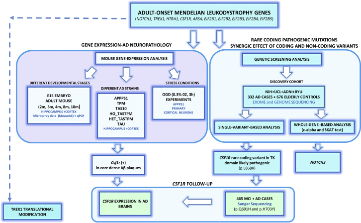
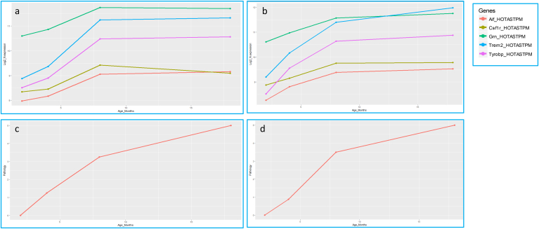
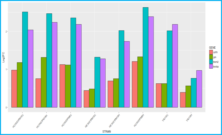
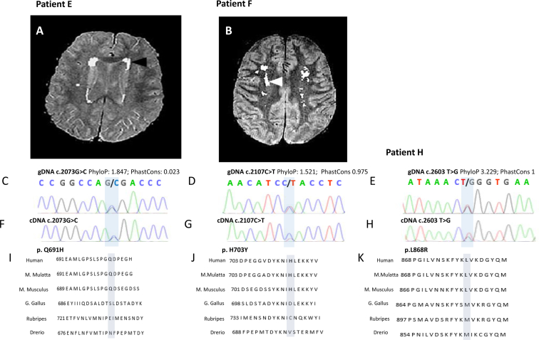
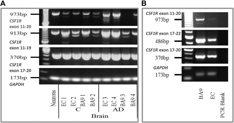
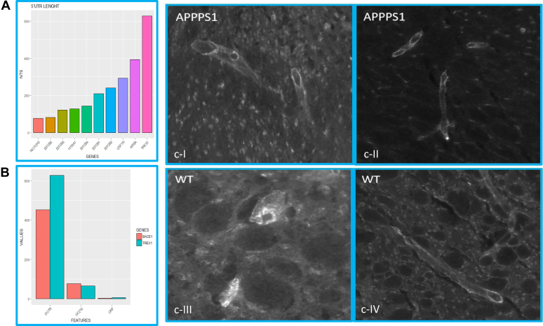
Mendelian adult-onset leukodystrophies are a spectrum of rare inherited progressive neurodegenerative disorders affecting the white matter of the central nervous system. Among these, cerebral autosomal dominant and recessive arteriopathy with subcortical infarcts and leukoencephalopathy, cerebroretinal vasculopathy, metachromatic leukodystrophy, hereditary diffuse leukoencephalopathy with spheroids, and vanishing white matter disease present with rapidly progressive dementia as dominant feature and are caused by mutations in NOTCH3, HTRA1, TREX1, ARSA, CSF1R, EIF2B1, EIF2B2, EIF2B3, EIF2B4, and EIF2B5, respectively. Given the rare incidence of these disorders and the lack of unequivocally diagnostic features, leukodystrophies are frequently misdiagnosed with common sporadic dementing diseases such as Alzheimer's disease (AD), raising the question of whether these overlapping phenotypes may be explained by shared genetic risk factors. To investigate this intriguing hypothesis, we have combined gene expression analysis (1) in 6 different AD mouse strains (APPPS1, HOTASTPM, HETASTPM, TPM, TAS10, and TAU) at 5 different developmental stages (embryo [E15], 2, 4, 8, and 18 months), (2) in APPPS1 primary cortical neurons under stress conditions (oxygen-glucose deprivation) and single-variant-based and single-gene-based (c-alpha test and sequence kernel association test (SKAT)) genetic screening in a cohort composed of 332 Caucasian late-onset AD patients and 676 Caucasian elderly controls. Csf1r was significantly overexpressed (log2FC > 1, adj. p-value < 0.05) in the cortex and hippocampus of aged HOTASTPM mice with extensive Aβ dense-core plaque pathology. We identified 3 likely pathogenic mutations in CSF1R TK domain (p.L868R, p.Q691H, and p.H703Y) in our discovery and validation cohort, composed of 465 AD and mild cognitive impairment (MCI) Caucasian patients from the United Kingdom. Moreover, NOTCH3 was a significant hit in the c-alpha test (adj p-value = 0.01). Adult-onset Mendelian leukodystrophy genes are not common factors implicated in AD. Nevertheless, our study suggests a potential pathogenic link between NOTCH3, CSF1R, and sporadic late-onset AD, which warrants further investigation.

Keywords: Alzheimer's disease; CSF1R; Mendelian leukodystrophies; NOTCH3.


Figures

Fig. 1
Fig. 1
Pipeline followed in the adult-onset leukodystrophy gene study. Abbreviations: AD, Alzheimer's disease; BYU, Brigham Young University; MCI, mild cognitive impairment; TK, tyrosine kinase; NIH, National Institutes of Health; UCL, University College London.
Fig. 2
Fig. 2
Log2-normalized expression of Csf1r, Grn, Trem2, Tyrobp, and Aif1 in HOTASTPM mice and related Aβ plaque pathology. (A–B) Log2-normalized expression of Csf1r, Grn, Trem2, Tyrobp and Aif1 in HOTASTPM mice (homozygous for the Swedish mutation APP p.K670N/M671L and PSEN1 p.M146V) at 4 different time points (2, 4, 8, and 18 months) in the hippocampus (A) and cortex (B) showing coexpression of the above genes. (C–D) Progression of AD pathology in hippocampus (C) and cortex (D), based on Aβ plaque density, in HOTASTPM mice at 4 different time points (2, 4, 8, and 18 months). Significant Csf1r, Grn, Trem2, Tyrobp, and Aif1 overexpression (Log2FC > 1, adj. p-value < 0.05) is detected at 8 months, in linear correlation with the most rapid and severe Aβ plaque deposition. Abbreviations: Aβ, amyloid beta; AD, Alzheimer's disease.
Fig. 3
Fig. 3
Log2FC of Csf1r, Grn, Trem2, and Tyrobp in different AD mouse strains during the most severe pathology, showing coexpression of Csf1r, Grn, Trem2, and Tyrobp. Particularly, Csf1r and Grn display the same overexpression pattern, which is, overall, 1/3 of Trem2 and Tyrobp upregulation. Abbreviations: C, cortex; H, hippocampus.
Fig. 4
Fig. 4
MRI scans, Sanger sequencing validation, and mutation domain conservation for patients E, F, and H. (A–B) Coronal T2-weighted MRI scan of patients E and F (for both taken 2y after onset of symptoms, aged 84y and 81y, respectively) showing symmetric patchy periventricular hyperintensities, mainly pronounced in the frontal lobes (A) and bilateral lesions localized to the centrum semiovale (B) (arrows). (C–E) Genomic DNA Sanger sequencing validation of CSF1R c.2073G>C, c.2107C>T, and c.2603 T>G mutations. (F–H) cDNA Sanger sequencing validation of c.2073G>C, c.2107C>T, and c.2603 T>G mutations. For patients F and H, cDNA sequence highlights a possible allelic imbalance, supporting the likely functional effect of gDNA c.2107C>T and gDNA c.2603 T>G mutations. (I–K) Conservation of p.Q691H, p. H703Y, and p.L868R in different species. PhastCons and PhyloP scores range between 0–1 and −14 to +6, respectively. For PhastCons, the closer to one, the more conserved; for PhyloP, conserved sites are assigned positive scores. Abbreviation: MRI, magnetic resonance imaging.
Fig. 5
Fig. 5
Tissue expression of CSF1R. (A) Relative CSF1R and GAPDH expression assessed by RT-PCR in postmortem brain RNA from normal control and AD individuals. (B) Expression of CSF1R transcripts and the house-keeping gene GAPDH in the EC and BA9 preassociation cortex (BA9) from patient H (c.2603T>G, p.L868R). RT-PCR for CSF1R exon 11 to 20, exon 11 to 19, exon 17 to 20, and exon 17 to 22 as indicated. Abbreviations: EC, entorhinal cortex; BA, Brodmann area; AD, Alzheimer’s disease; C, normal control; RT-PCR, Real Time PCR.
Supplementary Fig. S1
(A) Histological cortical section of APPPS1 mouse, 2 months of age, stained with Aβ oligomers (Ab126892). Rare Aβ oligomers are detected around small cortical vessels (B) and in the extracellular space, likely phagocytated by astrocytes (C), showing a very mild pathology and disease state. No Aβ plaques have been detected (data not shown).
Supplementary Fig. S2
(A) 5′UTR length of the studied adult-onset leukodystrophy genes. TREX1 presents the longest 5′UTR region (628nts). (B) TREX1 displays analogous 5′UTR features of BACE1, known to be subjected to translational control: (1) 5′UTR length (628 and 453 nts, respectively); (2) GC content (65% and 77%, respectively), and (3) predicted open reading frames (ORFs) (6 and 3, respectively). (CI–IV) Cortical histological sections of APPPS1 and WT mice (aged 2 months), stained with TREX1 antibody (Abnova 68191), which mainly binds to endothelial cells and to a lesser extent neurons. No macroscopical difference has been detected between APPPS1 and WT mice. Abbreviation: WT, wild type.

References

    1. Akiyama H., Nishimura T., Kondo H., Ikeda K., Hayashi Y., McGeer P.L. Expression of the receptor for macrophage colony stimulating factor by brain microglia and its upregulation in brains of patients with Alzheimer’s disease and amyotrophic lateral sclerosis. Brain Res. 1994;639:171–174. - PubMed
    1. Baba Y., Ghetti B., Baker M.C., Uitti R.J., Hutton M.L., Yamaguchi K., Bird T., Lin W., DeLucia M.W., Dickson D.W., Wszolek Z.K. Hereditary diffuse leukoencephalopathy with spheroids: clinical, pathologic and genetic studies of a new kindred. Acta Neuropathol. (Berl.) 2006;111:300–311. - PubMed
    1. Baker M., Mackenzie I.R., Pickering-Brown S.M., Gass J., Rademakers R., Lindholm C., Snowden J., Adamson J., Sadovnick A.D., Rollinson S., Cannon A., Dwosh E., Neary D., Melquist S., Richardson A., Dickson D., Berger Z., Eriksen J., Robinson T., Zehr C., Dickey C.A., Crook R., McGowan E., Mann D., Boeve B., Feldman H., Hutton M. Mutations in progranulin cause tau-negative frontotemporal dementia linked to chromosome 17. Nature. 2006;442:916–919. - PubMed
    1. Barber R., Scheltens P., Gholkar A., Ballard C., McKeith I., Ince P., Perry R., O’Brien J. White matter lesions on magnetic resonance imaging in dementia with Lewy bodies, Alzheimer's disease, vascular dementia, and normal aging. J. Neurol. Neurosurg. Psychiatry. 1999;67:66–72. - PMC - PubMed
    1. Boissé L., Islam O., Woulfe J., Ludwin S.K., Brunet D.G. Neurological picture. Hereditary diffuse leukoencephalopathy with neuroaxonal spheroids: novel imaging findings. J. Neurol. Neurosurg. Psychiatry. 2010;81:313–314. - PubMed

Publication types

MeSH terms

Substances