. 2018 May;39(5):817-823.
doi: 10.3174/ajnr.A5573. Epub 2018 Mar 15.

Localized Marked Elongation of the Distal Internal Carotid Artery with or without PHACE Syndrome: Segmental Dolichoectasia of the Distal Internal Carotid Artery

Affiliations

Localized Marked Elongation of the Distal Internal Carotid Artery with or without PHACE Syndrome: Segmental Dolichoectasia of the Distal Internal Carotid Artery

Z Y Jia et al. AJNR Am J Neuroradiol. 2018 May.

Abstract

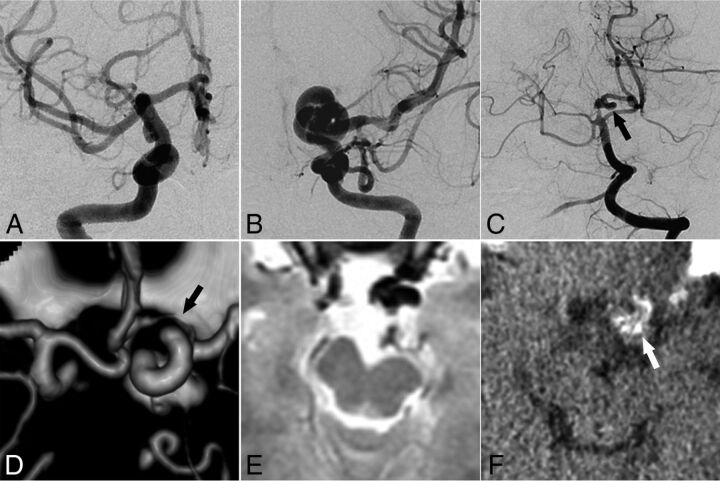
Background and purpose: Segmental intracranial dolichoectasia of the distal ICA is a feature of PHACE syndrome or a sporadic phenomenon. We evaluated the relationship between intracranial dolichoectasia of the distal ICA and PHACE syndrome and illustrated the characteristic radiologic findings of the lesions.

Materials and methods: Intracranial dolichoectasia of the distal ICA was identified in 20 patients at our institution from 2005 to 2016 through a review of diagnostic cerebral angiography results. All radiologic images were reviewed to determine the vascular morphologic dispositions around the distal ICA, including dysplasia, mural calcification, vessel wall enhancement, lumen narrowing, and aneurysm formation. Medical records were reviewed to determine the symptoms of PHACE syndrome. Subsequently, the correlation between radiologic findings and PHACE syndrome was assessed.

Results: In this cohort, which had a strong female predominance (male/female ratio= 2:18), intracranial dolichoectasia had a more ipsilateral vascular morphologic disposition. Mural calcification was detected more frequently in elderly patients, whereas vessel wall enhancement was detected more frequently in younger patients. Follow-up images showed a slow progression of the lesions. However, no significant differences in the vascular morphologic disposition and brain structural changes were observed between patients with (n = 11) and without (n = 9) PHACE syndrome.

Conclusions: The striking elongation and tortuosity of the distal ICA generally appeared to be a type of congenital lesion occurring early in embryogenesis as either a sporadic phenomenon or an arterial change associated with PHACE syndrome. Imaging findings revealed various mural abnormalities with a benign clinical course.


Figures

Fig 1.
Fig 1.
In addition to the 7 segments above, the carotid bulb to the PcomA origin, the segment of the ICA between the PcomA origin and ICA bifurcation before the MCA and anterior cerebral artery, is designated as the CS or distal ICA in adults. ILT indicates inferolateral trunk; MHT, meningohypophyseal trunk; VID, vidian artery; CT, caroticotympanic artery; CB, carotid bulb.
Fig 2.
Fig 2.
Representative images of ICDE in a patient without PHACE syndrome (patient 6, a 28-year-old woman). A and B, Bilateral ICA angiography shows a tangled arterial mass of the left distal ICA and no observable left A1 segment. C, Left vertebral artery angiography shows a dolichoectasia of the left P1 and P2 segments (arrow). D, CT angiography shows hypoplasia of the left A1 segment (arrow). E, T2-weighted image of the brain shows hypoplasia of the left midbrain. F, CT image of vessel wall calcification in the left distal ICA (arrow).
Fig 3.
Fig 3.
Evolution of vessel wall enhancement with aging in a patient with PHACE syndrome (patient 1, an 8-year-old girl). A and B, Left ICA angiography (anteroposterior and oblique views) reveals tortuosity in the left distal ICA with a small and long A1 segment. C and D, Noncontrast and contrast-enhanced vessel wall imaging at 8 years of age demonstrates vessel wall enhancement and slight vessel wall thickening (arrow in D). E and F, Vessel wall imaging at 12 years of age shows a similar vessel wall enhancement pattern (arrow in F).
Fig 4.
Fig 4.
Vessel wall enhancement at the stenotic segment in a patient with possible PHACE syndrome (patient 5, a 23-year-old woman). A and B, Left ICA angiography and TOF-MRA reveal dolichoectasia of the left distal ICA, left PcomA (arrow), and left AchoA (curved arrow). C–E, Section images reveal the vessel wall thickening and enhancing patterns of each corresponding stenotic segment (arrowheads indicate the vessel wall thickening and asterisks indicate vessel lumen; C, sagittal plane; D and E, axial plane). P indicates posterior; R, right.
Fig 5.
Fig 5.
Vessel wall enhancement at the aneurysm wall and the parent artery wall in a patient without PHACE syndrome (patient 11, a 45-year-old woman). A–C, Cerebral angiography shows dolichoectasia in the right distal ICA and basilar artery. D, 3D-DSA shows dolichoectasia in the right distal ICA and right PcomA as well as the formation of multiple aneurysms in the right PcomA (arrow). E and F, Non-contrast-enhanced and contrast-enhanced vessel wall imaging shows enhancement of the aneurysm wall (arrows in F) and the parent artery wall (curved arrow in F).

Comment in

Similar articles

Cited by

References

    1. Burch EA, Garzon MC, Parikh A, et al. . A 65-year-old woman diagnosed with PHACE syndrome. Pediatr Dermatol 2013;30:e153–56 10.1111/pde.12043 - DOI - PubMed
    1. Baccin CE, Krings T, Alvarez H, et al. . A report of two cases with dolichosegmental intracranial arteries as a new feature of PHACES syndrome. Childs Nerv Syst 2007;23:559–67 10.1007/s00381-006-0247-8 - DOI - PubMed
    1. Metry DW, Dowd CF, Barkovich AJ, et al. . The many faces of PHACE syndrome. J Pediatr 2001;139:117–23 10.1067/mpd.2001.114880 - DOI - PubMed
    1. Rossi A, Bava GL, Biancheri R, et al. . Posterior fossa and arterial abnormalities in patients with facial capillary haemangioma: presumed incomplete phenotypic expression of PHACES syndrome. Neuroradiology 2001;43:934–40 10.1007/s002340100594 - DOI - PubMed
    1. McLaughlin N, Raychev R, Duckwiler G, et al. . Pure arterial malformation of the posterior cerebral artery: importance of its recognition. J Neurosurg 2013;119:655–60 10.3171/2013.4.JNS121374 - DOI - PubMed

Supplementary concepts

LinkOut - more resources