. 2018 Jun 1;78(11):2966-2977.
doi: 10.1158/0008-5472.CAN-17-2269. Epub 2018 Mar 15.

Mutant IDH1 Cooperates with ATRX Loss to Drive the Alternative Lengthening of Telomere Phenotype in Glioma

Affiliations

Mutant IDH1 Cooperates with ATRX Loss to Drive the Alternative Lengthening of Telomere Phenotype in Glioma

Joydeep Mukherjee et al. Cancer Res. .

Abstract

A subset of tumors use a recombination-based alternative lengthening of telomere (ALT) pathway to resolve telomeric dysfunction in the absence of TERT. Loss-of-function mutations in the chromatin remodeling factor ATRX are associated with ALT but are insufficient to drive the process. Because many ALT tumors express the mutant isocitrate dehydrogenase IDH1 R132H, including all lower grade astrocytomas and secondary glioblastoma, we examined a hypothesized role for IDH1 R132H in driving the ALT phenotype during gliomagenesis. In p53/pRb-deficient human astrocytes, combined deletion of ATRX and expression of mutant IDH1 were sufficient to create tumorigenic cells with ALT characteristics. The telomere capping complex component RAP1 and the nonhomologous DNA end joining repair factor XRCC1 were each downregulated consistently in these tumorigenic cells, where their coordinate reexpression was sufficient to suppress the ALT phenotype. RAP1 or XRCC1 downregulation cooperated with ATRX loss in driving the ALT phenotype. RAP1 silencing caused telomere dysfunction in ATRX-deficient cells, whereas XRCC1 silencing suppressed lethal fusion of dysfunctional telomeres by allowing IDH1-mutant ATRX-deficient cells to use homologous recombination and ALT to resolve telomeric dysfunction and escape cell death. Overall, our studies show how expression of mutant IDH1 initiates telomeric dysfunction and alters DNA repair pathway preferences at telomeres, cooperating with ATRX loss to defeat a key barrier to gliomagenesis.Significance: Studies show how expression of mutant IDH1 initiates telomeric dysfunction and alters DNA repair pathway preferences at telomeres, cooperating with ATRX loss to defeat a key barrier to gliomagenesis and suggesting new therapeutic options to treat low-grade gliomas. Cancer Res; 78(11); 2966-77. ©2018 AACR.


Conflict of interest statement

Disclosure of Potential Conflicts of Interest

No potential conflicts of interest were disclosed.

Figures

Figure 1.
Figure 1.
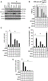
Mutant IDH1 cooperates with ATRX loss to stimulate ALT. A, PCR-based analysis of DNA from E6E7-expressing human astrocytes (Cont) exogenously expressing mutant IDH1 (+mutIDH1), subjected to CRISPR-based homozygous deletion of ATRX (ATRX KO-1 and ATRX KO-2), or both yields a 700-bp product in cells having undergone successful homozygous deletion of ATRX exons 7–9. B, Western blot verification of ATRX, mutant IDH1, and β-actin expression in cells in A. C, TRAP analysis of telomerase activity in cells in A and in control E6E7 astrocytes exogenously expressing hTERT (leftmost lane, closed circle). D, Quantitation of the percentage of cells containing ≤1 (open box) or 2–4 (closed box) APBs per cell (bottom) based on IHC colocalization (yellow foci) of PML (red) with TRF2 (green) signal (top) in DAPI-stained (blue) positive-control GM847 ALT cells and the ATRX-KO1–based cells in A. N ≥ 200 cells per group. E, Quantitation of the percentage of chromosomes with T-SCE (bottom) based on fluorescence colocalization of leading (red)- and lagging (green)–strand telomeric probes (top) in >50 cells per ATRX-KO1-based cells in A. F, Quantitation of C-circle DNA following amplification of varying amounts of genomic DNA from GM847 and ATRX KO-1–based cells in A in reactions with (+) or without (−) phi29 polymerase that were spotted (dotted areas) and hybridized to a telomeric G strand-specific probe. G, Number of colonies (>100 cells) that arose 28 days following plating of the ATRX KO-1–based cells in A in soft agar. Except where noted, all values were derived from three independent experiments. *, P < 0.05.
Figure 2.
Figure 2.
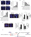
Mutant IDH1 expression is associated with downregulation of RAP1 and XRCC1. A, Triplicate quantitative PCR analysis of the levels of select transcripts encoding proteins involved in telomere regulation in control cells (E6E7 astrocytes) expressing mutant IDH1, subjected to CRISPR-based deletion of ATRX, or both. B, Western blot analysis of RAP1, XRCC1, DNA ligase 3 (Lig3), and β-actin protein levels in the cells in A and in BT142, SF10602, and MGG119 IDH1-mutant ALT glioma cells. C, The Cancer Genome Atlas–based analysis of XRCC1 and RAP1 mRNA levels in LGA, lower-grade mixed oligoastroglioma (LOA), and GBM relative to normal brain controls (1.0 on the y-axis). *, P < 0.05.
Figure 3.
Figure 3.
Forced overexpression of RAP1 and/or XRCC1 suppresses mutant IDH1-mediated ALT phenotype in genetically modified human astrocytes. A, Western blot verification of ATRX, mutant IDH1, RAP1, XRCC1, and β-actin protein levels in ATRX KO-1 astrocytes, and ATRX KO-1 astrocytes exogenously expressing mutant IDH1 plus ATRX, RAP1, XRCC1, or both RAP1 and XRCC1. B, Quantitation of the percentage of cells from A containing ≤1 (open box) or 2–4 (closed box) APBs per cell. N ≥ 200 cells per group. ATRX (−), CRISPR-based deletion of ATRX, ATRX (+), exogenous re-introduction of ATRX. C, Percentage of chromosomes with T-SCE in cells from A. D, PCR-based quantification of C-circle signal (relative to that in positive control GM847 ALT fibroblasts) following phi29-mediated amplification of genomic DNA (30 ng) from cells in A. E, Number of colonies (>100 cells) that arose 28 days following plating of the cells in A. Except where noted, all values were derived from three independent experiments. *, P < 0.05.
Figure 4.
Figure 4.
Forced overexpression of RAP1 and/or XRCC1 suppresses mutant IDH1–mediated ALT phenotype in ATRX-deficient, mutant IDH1, ALT MGG119 xenograft cells. A, Western blot verification of ATRX, RAP1, XRCC1, and β-actin protein levels in MGG119 cells, and the same cells exogenously expressing ATRX, RAP1, XRCC1, or both RAP1 and XRCC1. B, Quantitation of the percentage of cells containing ≤1 (open box) or 2–4 (closed box) APBs per cell in >200 cells per each group described in A. +/− symbols denote the exogenous introduction of ATRX, RAP1, or XRCC1 (+), or of blank vector (−). C, Percentage of chromosomes with T-SCE in cells in A. D, PCR-based quantification of C-circle signal (relative to that in positive control GM847 ALT fibroblasts) following phi29-mediated amplification of genomic DNA (30 ng) from cells in A. E, Viability of cells in A measured by trypan blue exclusion. F, Number of colonies (>100 cells) that arose 28 days following plating of the cells in A. Except where noted, all values were derived from three independent experiments. *, P < 0.05.
Figure 5.
Figure 5.
siRNA-mediated suppression of RAP1 and/or XRCC1 substitutes for mutant IDH1 in driving the ALT phenotype. A, Western blot verification of IDH1, RAP1, XRCC1, and β-actin protein levels in ATRX KO-1 astrocytes transfected with scrambled siRNA or pooled siRNAs targeting RAP1, XRCC1, or both. B, Quantitation of the percentage of cells containing ≤1 (open box) or 2–4 (closed box) APBs per cell in >200 cells per each group described in A. C, Percentage of chromosomes with T-SCE from ATRX KO cells in A and also expressing blank (−) or mutant IDH1-encoding construct (+). D, Quantitation of C-circle DNA in cells from A. E, Number of colonies (>100 cells) that arose 28 days following plating of the cells in D. Except where noted, all values were derived from three independent experiments.*, P < 0.05.
Figure 6.
Figure 6.
Mutant IDH1-mediated downregulation of RAP1 and XRCC1 initiates telomere dysfunction and alters telomeric damage repair. A, Quantitation (bottom) of the percentage of control E6E7-expressing human astrocytes expressing mutant IDH1 (IDH1 mut+), subjected to CRISPR-based ATRX knockout, (ATRX KO-1) (ATRX−), or both and containing >2 TIFs per cell (TIF positive, yellow foci, top) based on IHC colocalization of γH2AX (red) with TRF2 (green) signal in >50 cells per group. B, Quantitation of the percentage of TIF-positive mutant IDH1/ATRX KO-1 cells following introduction of blank constructs or constructs encoding RAP1, XRCC1, or both. C, Quantitation of the percentage of TIF-positive IDH1 WT/ATRX KO-1 cells following introduction of scrambled siRNA (−) or siRNA targeting RAP1 and/or XRCC1 (+). D and E, Quantitation of the viability (right, dark bars) and the percentage of mutant IDH1/ATRX KO-1 (D) or MGG119 (E) cells containing ≥1 fused chromosome (right, light bars) following introduction of blank constructs or constructs encoding XRCC1, based on fluorescence microscopy analysis of >50 cells per each group (left). F, Schematic of the basis for mutant IDH1–mediated control of ALT. Except where noted, all values were derived from three independent experiments. *, P < 0.05.

References

    1. Greider CW. Telomerase and telomere-length regulation: lessons from small eukaryots to mammals. Cold Spring Harb Symp Quant Biol 1993; 58:719–23. - PubMed
    1. Muraki K, Nyhan K, Han L, Murnane J. Mechanisms of telomere loss and their consequences for chromosome instability. Front Oncol 2012;2:135. - PMC - PubMed
    1. Palm W, de Lange T. How shelterin protects mammalian telomeres. Annu Rev Genet 2008;42:301–34. - PubMed
    1. Lazzerini-Denchi E, Sfeir A. Stop pulling my strings-what telomeres taught us about the DNA damage response. Nat Rev 2016;17:364–78. - PMC - PubMed
    1. Eckel-Passow JE, Lachance DH, Molinaro AM, Walsh KM, Decker PA, Sicotte H, et al. Glioma groups based on 1p/19q, IDH, and TERT promoter mutations in tumors. N Engl J Med 2015;372:2499–508. - PMC - PubMed

Publication types