Distinct protein signatures of acute myeloid leukemia bone marrow-derived stromal cells are prognostic for patient survival
- PMID: 29545342
- PMCID: PMC5927978
- DOI: 10.3324/haematol.2017.172429
Distinct protein signatures of acute myeloid leukemia bone marrow-derived stromal cells are prognostic for patient survival
Abstract
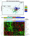
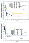
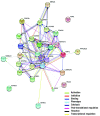
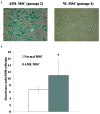
Mesenchymal stromal cells (MSC) support acute myeloid leukemia (AML) cell survival in the bone marrow (BM) microenvironment. Protein expression profiles of AML-derived MSC are unknown. Reverse phase protein array analysis was performed to compare expression of 151 proteins from AML-MSC (n=106) with MSC from healthy donors (n=71). Protein expression differed significantly between the two groups with 19 proteins over-expressed in leukemia stromal cells and 9 over-expressed in normal stromal cells. Unbiased hierarchical clustering analysis of the samples using these 28 proteins revealed three protein constellations whose variation in expression defined four MSC protein expression signatures: Class 1, Class 2, Class 3, and Class 4. These cell populations appear to have clinical relevance. Specifically, patients with Class 3 cells have longer survival and remission duration compared to other groups. Comparison of leukemia MSC at first diagnosis with those obtained at salvage (i.e. relapse/refractory) showed differential expression of 9 proteins reflecting a shift toward osteogenic differentiation. Leukemia MSC are more senescent compared to their normal counterparts, possibly due to the overexpressed p53/p21 axis as confirmed by high β-galactosidase staining. In addition, overexpression of BCL-XL in leukemia MSC might give survival advantage under conditions of senescence or stress and overexpressed galectin-3 exerts profound immunosuppression. Together, our findings suggest that the identification of specific populations of MSC in AML patients may be an important determinant of therapeutic response.
Copyright © 2018 Ferrata Storti Foundation.
Figures


References
-
- Barcellos-de-Souza P, Gori V, Bambi F, Chiarugi P. Tumor microenvironment: bone marrow-mesenchymal stem cells as key players. Biochim Biophys Acta. 2013;1836(2):321–335. - PubMed
-
- da Silva Meirelles L, Chagastelles PC, Nardi NB. Mesenchymal stem cells reside in virtually all post-natal organs and tissues. J Cell Sci. 2006;119(Pt 11):2204–2213. - PubMed
Publication types
MeSH terms
Substances
Grants and funding
LinkOut - more resources
Full Text Sources
Other Literature Sources
Medical
Research Materials
Miscellaneous

