. 2018 Jun;103(6):959-971.
doi: 10.3324/haematol.2017.183202. Epub 2018 Mar 15.

Cytokines increase engraftment of human acute myeloid leukemia cells in immunocompromised mice but not engraftment of human myelodysplastic syndrome cells

Affiliations

Cytokines increase engraftment of human acute myeloid leukemia cells in immunocompromised mice but not engraftment of human myelodysplastic syndrome cells

Maria Krevvata et al. Haematologica. 2018 Jun.

Abstract

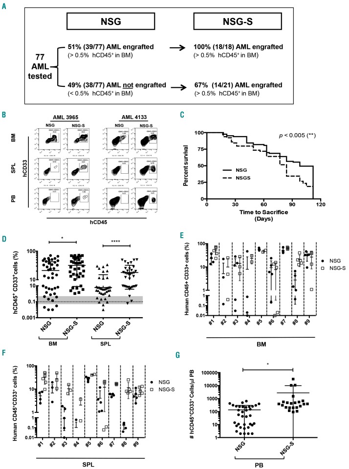
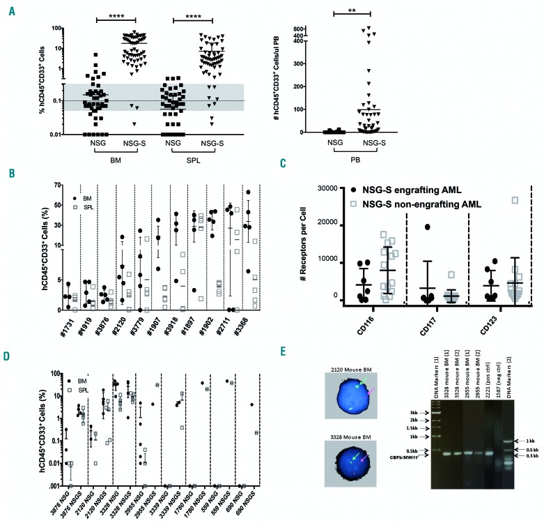
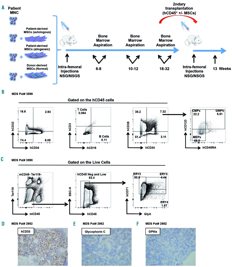
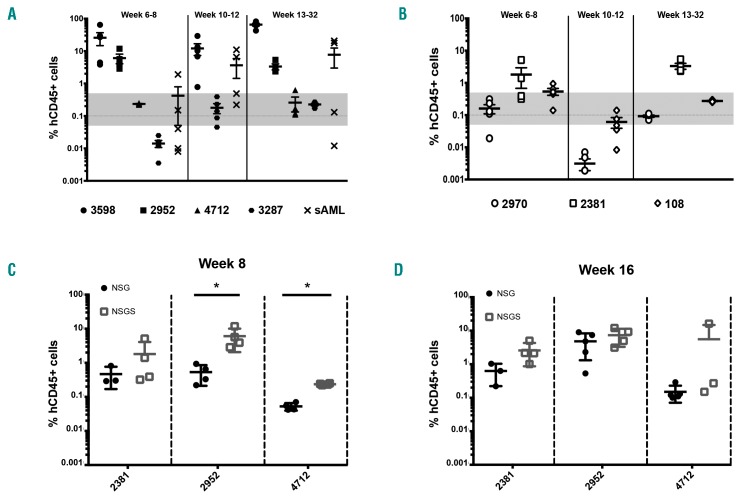
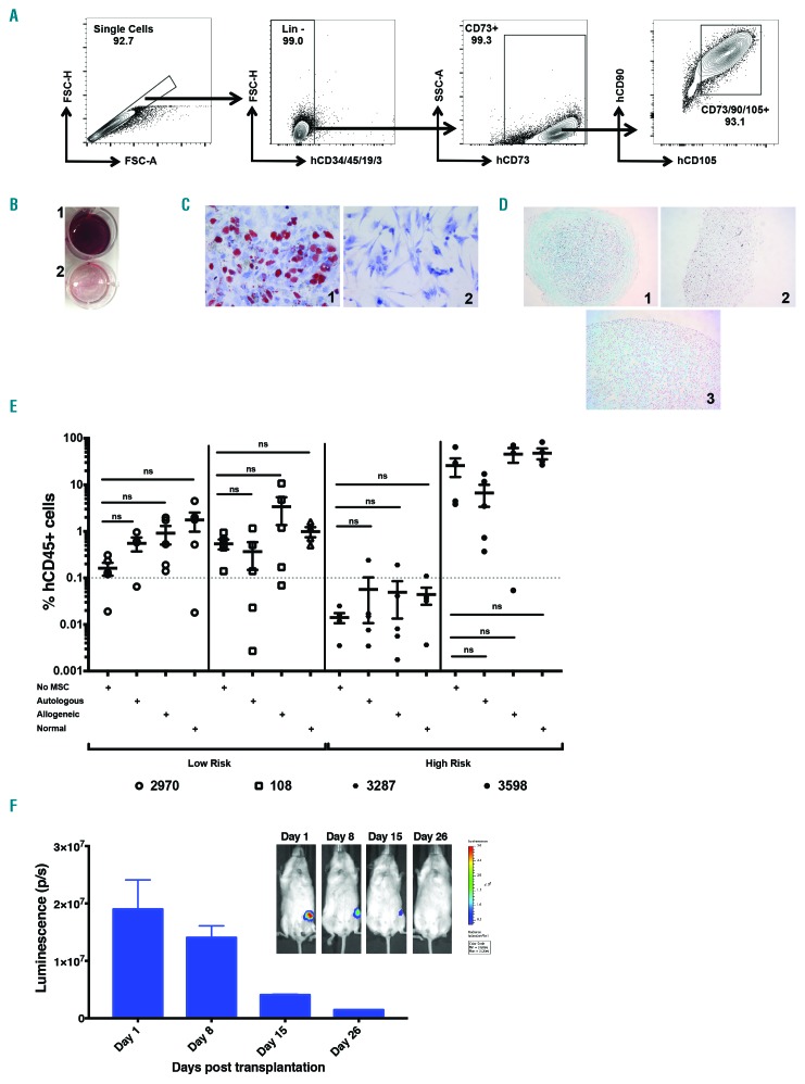
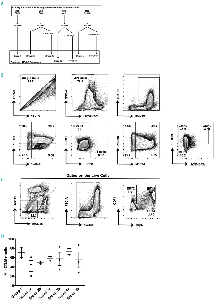
Patient-derived xenotransplantation models of human myeloid diseases including acute myeloid leukemia, myelodysplastic syndromes and myeloproliferative neoplasms are essential for studying the biology of the diseases in pre-clinical studies. However, few studies have used these models for comparative purposes. Previous work has shown that acute myeloid leukemia blasts respond to human hematopoietic cytokines whereas myelodysplastic syndrome cells do not. We compared the engraftment of acute myeloid leukemia cells and myelodysplastic syndrome cells in NSG mice to that in NSG-S mice, which have transgene expression of human cytokines. We observed that only 50% of all primary acute myeloid leukemia samples (n=77) transplanted in NSG mice provided useful levels of engraftment (>0.5% human blasts in bone marrow). In contrast, 82% of primary acute myeloid leukemia samples engrafted in NSG-S mice with higher leukemic burden and shortened survival. Additionally, all of 5 injected samples from patients with myelodysplastic syndrome showed persistent engraftment on week 6; however, engraftment was mostly low (<2%), did not increase over time, and was only transiently affected by the use of NSG-S mice. Co-injection of mesenchymal stem cells did not enhance human myelodysplastic syndrome cell engraftment. Overall, we conclude that engraftment of acute myeloid leukemia samples is more robust compared to that of myelodysplastic syndrome samples and unlike those, acute myeloid leukemia cells respond positively to human cytokines, whereas myelodysplastic syndrome cells demonstrate a general unresponsiveness to them.


Figures

Figure 1.
Figure 1.
NSG-S mice showed higher and more rapid engraftment of primary patients’ acute myeloid leukemia (AML) samples than did NSG mice. (A) Thirty-nine of the 77 AML samples screened were engrafted (hCD45+CD33+ blasts > 0.5% in mouse bone marrow) in NSG mice. All 39 AML engrafted in NSG mice also engrafted in NSG-S mice. Thirty-eight AML samples did not engraft in NSG, and approximately two-thirds of these engrafted in NSG-S mice. (B) Representative flow cytometry plots of human hCD45+CD33+ leukemia cells from bone marrow (BM), spleen (SPL) and peripheral blood (PB) samples from NSG and NSG-S mice. (C) Kaplan-Meier curves show the time to sacrifice mice for all 77 AML samples. (D) Leukemia burden of 9 AML engrafted in both NSG and NSG-S mice which were sacrificed at the same time was assessed by % of human hCD45+CD33+ cells in mouse BM and SPL. Each symbol represents one mouse. (E and F) Individual mouse BM and SPL leukemia burdens of the 9 AML engrafters are shown. Solid symbols represent data from NSG mice, and empty symbols represent data from NSG-S mice. (G) Elevated levels of leukemia burden (% hCD45+CD33+ cells) were seen in NSG-S mice.
Figure 2.
Figure 2.
NSG-S mice enhanced the engraftment of human patients’ acute myeloid leukemia (AML) samples more than NSG mice (non-NSG engrafted patients’ samples were used). (A) Levels of hCD45+CD33+ leukemia blasts in bone marrow (BM), spleen (SPL) and peripheral blood (PB) of NSG or NSG-S mice (n=14) injected with the same number of AML cells. (B) Individual mouse BM and SPL leukemia burdens of 11 out of the 14 mice from (A) are shown. (C) Receptor densities (CD116, CD117 and CD123) on AML cells of the 7 non-NSG-S engrafters and 14 NSG-S engrafters were assessed as number of receptors per cell. (D) Establishment of 8 inv(16) AML patient-derived xenotransplant models in NSG-S mice. (E) The chromosomal abnormalities for inv(16) were confirmed in the BM of engrafted NSG-S mice by fluorescent in situ hybridization (left panels) and breakpoint reverse transcriptase polymerase chain reaction (right panel).
Figure 3.
Figure 3.
Myelodysplastic syndromes (MDS) cells engraft in NSG-S mice. (A) Schematic representation of experimental set up. Patient-derived bone marrow mononuclear cells (MNC) alone or in combination with mesenchymal stem cells (MSC) were intrafemorally injected in NSG-S mice. Characteristic gating strategy for analyzing the (B) myeloid (CD33), lymphoid (CD19, CD3), progenitor (CD45RA, CD123, CD38) and (C) erythroid (GlyA) subpopulations found in the bone marrow of mice engrafted with patients’ bone marrow MNC samples. Immunostainings of (D) hCD33, (E) glycophorin C and (F) GPIIIa of decalcified bone marrow sections from sternums of mice engrafted with human MDS.
Figure 4.
Figure 4.
Most myeloproliferative syndrome (MDS) samples do not show sustained engraftment and human cytokines moderately enhance engraftment. Time course representation of the percentages of hCD45+ cells in the bone marrow of xenografted NSG-S mice injected with (A) high risk and (B) low risk human MDS patients’ mononuclear cells. The gray line corresponds to the 0.1% threshold used in the study and the gray zone reflects a broader area of uncertainty of engraftment. Bone marrow mononuclear cells from patients diagnosed with MDS were intrafemorally injected into NSG and NSG-S mice in a strain comparison experiment. Percentage of hCD45+ cells found in the murine bone marrow was evaluated at (C) eight and (D) 16 weeks after transplantation.
Figure 5.
Figure 5.
Myelodysplastic syndrome (MDS) cell engraftment is not affected by mesenchymal stem cells (MSC). (A) Representative phenotypical characterization of the ex vivo-expanded human MSC used in the transplantation experiments. Trilineage in vitro differentiation of ex vivo expanded MDS patient MSC into (B) osteoblasts, (C) adipocytes, and (D) chondrocytes. [(1) Differentiated, (2) Undifferentiated, (3) Positive Control] (E) Bone marrow human mononuclear cells were intrafemorally transplanted into NSG-S mice alone or in combination with ex vivo-expanded MSC isolated from various origins. Percentages of hCD45+ cells present in the murine bone marrow at 6–8 weeks post transplantation. Red line at 0.1% represents the positive engraftment threshold level. Comparisons showed no significant statistical differences between the four groups. (F) NSG mice intrafemorally transplanted with labeled MSC-green fluorescent protein-luciferase. Bioluminescence levels were measured in vivo at the timepoints indicated.
Figure 6.
Figure 6.
Myelodysplastic syndrome-initiating cells can be maintained in NSG-S mice. (A) Schematic representation of experimental set up for secondary intrafemoral transplantations. (Β) Percentages of hCD45+ cells at 13 weeks after transplantation. Characteristic gating strategy for analyzing the (C) myeloid (CD33), lymphoid (CD19, CD3), progenitor (CD45RA, CD123, CD38) and (D) erythroid (GlyA) subpopulations found in the bone marrow of mice engrafted with hCD45+ cells isolated from primary mice.

Comment in

References

    1. Tallman MS, Gilliland DG, Rowe JM. Drug therapy for acute myeloid leukemia. Blood. 2005;106(4):1154–1163. - PubMed
    1. Walter MJ, Shen D, Ding L, et al. Clonal architecture of secondary acute myeloid leukemia. N Engl J Med. 2012;366(12):1090–1098. - PMC - PubMed
    1. Will B, Zhou L, Vogler TO, et al. Stem and progenitor cells in myelodysplastic syndromes show aberrant stage-specific expansion and harbor genetic and epigenetic alterations. Blood. 2012;120(10):2076–2086. - PMC - PubMed
    1. Woll PS, Kjällquist U, Chowdhury O, et al. Myelodysplastic syndromes are propagated by rare and distinct human cancer stem cells in vivo. Cancer Cell. 2014;25(6):794–808. - PubMed
    1. Schofield R. The relationship between the spleen colony-forming cell and the haemopoietic stem cell. Blood Cells. 1978;4(1–2):7–25. - PubMed

Publication types

MeSH terms