

Helminth infection protects against high fat diet-induced obesity via induction of alternatively activated macrophages
- PMID: 29545532
- PMCID: PMC5854586
- DOI: 10.1038/s41598-018-22920-7
Helminth infection protects against high fat diet-induced obesity via induction of alternatively activated macrophages
Abstract
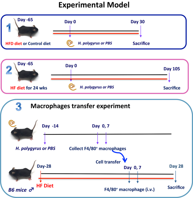
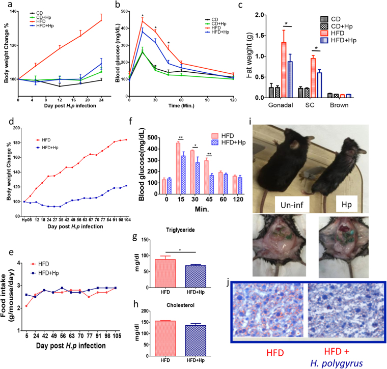
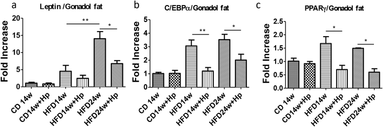
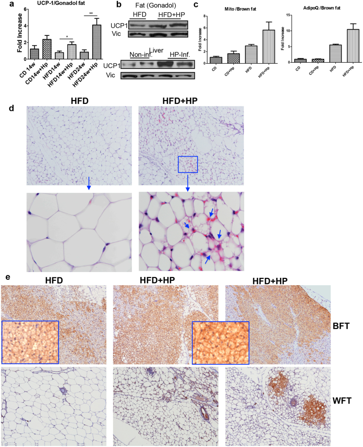
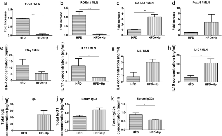
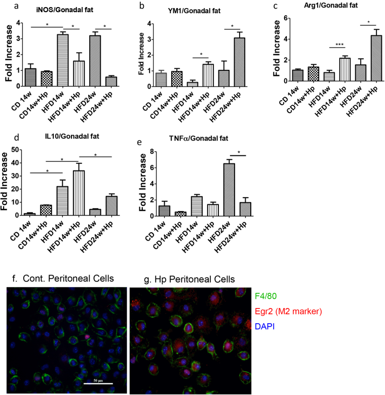
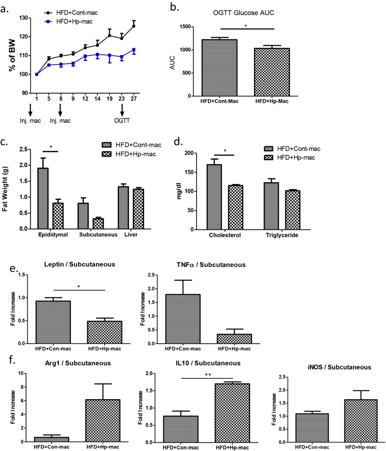
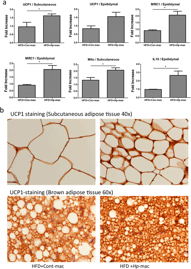
Epidemiological studies indicate an inverse correlation between the prevalence of the so-called western diseases, such as obesity and metabolic syndrome, and the exposure to helminths. Obesity, a key risk factor for many chronic health problems, is rising globally and is accompanied by low-grade inflammation in adipose tissues. The precise mechanism by which helminths modulate metabolic syndrome and obesity is not fully understood. We infected high fat diet (HFD)-induced obese mice with the intestinal nematode parasite Heligmosomoides polygyrus and observed that helminth infection resulted in significantly attenuated obesity. Attenuated obesity corresponded with marked upregulation of uncoupling protein 1 (UCP1), a key protein involved in energy expenditure, in adipose tissue, suppression of glucose and triglyceride levels, and alteration in the expression of key genes involved in lipid metabolism. Moreover, the attenuated obesity in infected mice was associated with enhanced helminth-induced Th2/Treg responses and M2 macrophage polarization. Adoptive transfer of helminth-stimulated M2 cells to mice that were not infected with H. polygyrus resulted in a significant amelioration of HFD-induced obesity and increased adipose tissue browning. Thus, our results provide evidence that the helminth-dependent protection against obesity involves the induction of M2 macrophages.
Conflict of interest statement
The authors declare no competing interests.
Figures
Similar articles
-
Helminth-Induced and Th2-Dependent Alterations of the Gut Microbiota Attenuate Obesity Caused by High-Fat Diet.Cell Mol Gastroenterol Hepatol. 2020;10(4):763-778. doi: 10.1016/j.jcmgh.2020.06.010. Epub 2020 Jul 3. Cell Mol Gastroenterol Hepatol. 2020. PMID: 32629118 Free PMC article.
-
IL-33 is essential to prevent high-fat diet-induced obesity in mice infected with an intestinal helminth.Parasite Immunol. 2020 Sep;42(9):e12700. doi: 10.1111/pim.12700. Epub 2020 Jun 16. Parasite Immunol. 2020. PMID: 32027755
-
Macrophage Migration Inhibitory Factor (MIF) Is Essential for Type 2 Effector Cell Immunity to an Intestinal Helminth Parasite.Front Immunol. 2019 Oct 24;10:2375. doi: 10.3389/fimmu.2019.02375. eCollection 2019. Front Immunol. 2019. PMID: 31708913 Free PMC article.
-
Immunity to the model intestinal helminth parasite Heligmosomoides polygyrus.Semin Immunopathol. 2012 Nov;34(6):829-46. doi: 10.1007/s00281-012-0347-3. Epub 2012 Oct 11. Semin Immunopathol. 2012. PMID: 23053394 Free PMC article. Review.
-
Interactions between macrophages and helminths.Parasite Immunol. 2020 Jul;42(7):e12717. doi: 10.1111/pim.12717. Epub 2020 May 3. Parasite Immunol. 2020. PMID: 32249432 Review.
Cited by
-
Anti-obesity effects by parasitic nematode (Trichinella spiralis) total lysates.Front Cell Infect Microbiol. 2024 Jan 8;13:1285584. doi: 10.3389/fcimb.2023.1285584. eCollection 2023. Front Cell Infect Microbiol. 2024. PMID: 38259965 Free PMC article.
-
Suppression of Obesity by an Intestinal Helminth through Interactions with Intestinal Microbiota.Infect Immun. 2019 May 21;87(6):e00042-19. doi: 10.1128/IAI.00042-19. Print 2019 Jun. Infect Immun. 2019. PMID: 30962398 Free PMC article.
-
Helminth infection modulates systemic pro-inflammatory cytokines and chemokines implicated in type 2 diabetes mellitus pathogenesis.PLoS Negl Trop Dis. 2020 Mar 3;14(3):e0008101. doi: 10.1371/journal.pntd.0008101. eCollection 2020 Mar. PLoS Negl Trop Dis. 2020. PMID: 32126084 Free PMC article.
-
Resident TH2 cells orchestrate adipose tissue remodeling at a site adjacent to infection.Sci Immunol. 2022 Oct 21;7(76):eadd3263. doi: 10.1126/sciimmunol.add3263. Epub 2022 Oct 14. Sci Immunol. 2022. PMID: 36240286 Free PMC article.
-
The Adipose Tissue Macrophages Central to Adaptive Thermoregulation.Front Immunol. 2022 Apr 12;13:884126. doi: 10.3389/fimmu.2022.884126. eCollection 2022. Front Immunol. 2022. PMID: 35493493 Free PMC article. Review.
References
Publication types
MeSH terms
Substances
Grants and funding
LinkOut - more resources
Full Text Sources
Other Literature Sources
Medical
Research Materials
