. 2018 Mar 15;8(1):4632.
doi: 10.1038/s41598-018-22855-z.

Secretome analysis of in vitro aged human mesenchymal stem cells reveals IGFBP7 as a putative factor for promoting osteogenesis

Affiliations

Secretome analysis of in vitro aged human mesenchymal stem cells reveals IGFBP7 as a putative factor for promoting osteogenesis

Arantza Infante et al. Sci Rep. .

Abstract

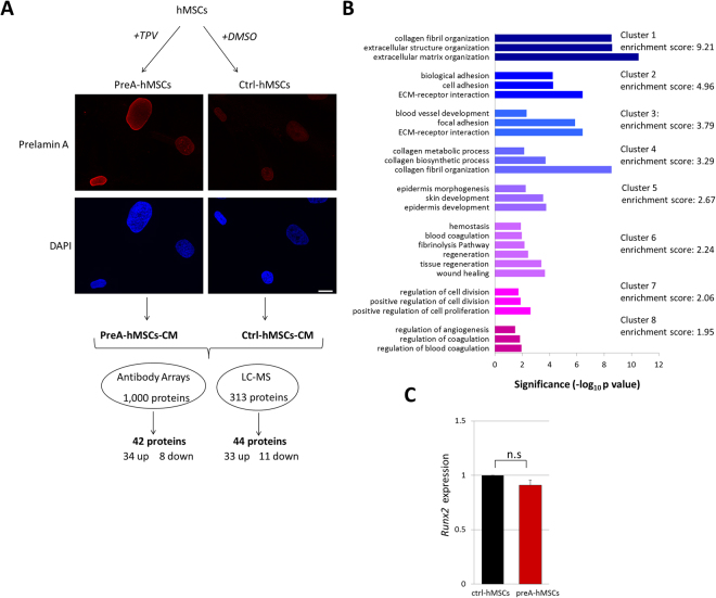
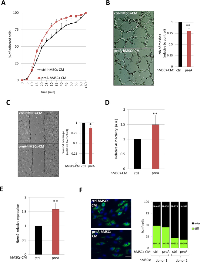
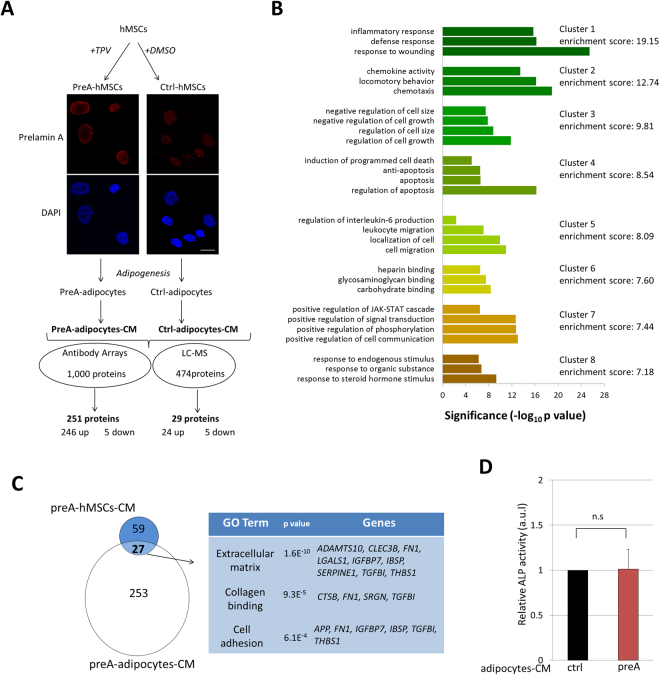
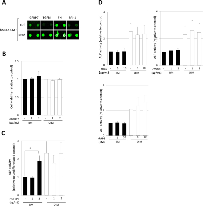
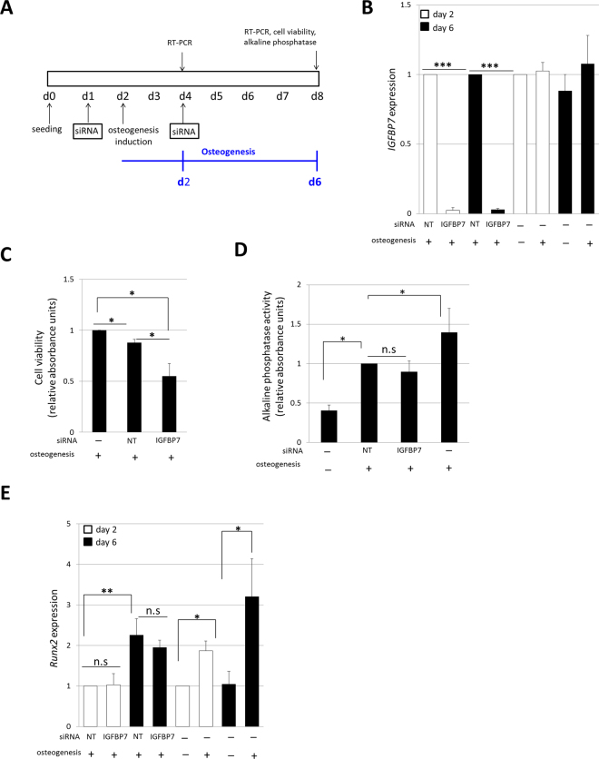
Aging is a complex biological process, which involves multiple mechanisms with different levels of regulation. Senescent cells are known to secrete senescence-associated proteins, which exert negative influences on surrounding cells. Mesenchymal stem cells (MSCs), the common progenitors for bone, cartilage and adipose tissue (which are especially affected tissues in aging), are known to secrete a broad spectrum of biologically active proteins with both paracrine and autocrine functions in many biological processes. In this report, we have studied the secreted factors (secretome) from human MSCs (hMSCs) and hMSCs-derived adipocytes which were induced to accumulate prelamin A, the immature form of the nuclear lamina protein called Lamin A, known to induce premature aging syndromes in humans and in murine models. Proteomic analysis from two different techniques, antibody arrays and LS-MS, showed that prelamin A accumulation in hMSCs promotes the differential secretion of factors previously identified as secreted by hMSCs undergoing osteogenesis. Moreover, this secretome was able to modulate osteogenesis of normal hMSCs in vitro. Finally, we found that one of the overexpressed secreted factors of this human aging in vitro stem cell model, IGFBP-7, is an osteogenic factor, essential for the viability of hMSCs during osteogenesis.


Conflict of interest statement

The authors declare no competing interests.

Figures

Figure 1
Figure 1
Analysis of preA-hMSCs-CM reveals altered secretion of proteins related to extracellular matrix, cell adhesion, angiogenesis and wound healing. (A) Schematic overview of hMSCs treatment to induce prelamin A accumulation. From each hMSCs line hMSCs accumulating prelamin A (preA-hMSCs) and control hMSCs (ctrl-hMSCs) were obtained in parallel. Prelamin A accumulation at the nuclear envelope was confirmed by confocal microscopy (red: prelamin A, blue: DAPI). Scale bar = 10 µm. Conditioned media from preA-hMSCs and ctrl-hMSCs were collected and subjected to proteomic analysis. Two independent hMSCs lines were use to obtain conditioned media in the case of antibody arrays (n = 2) and 4 independent hMSCs lines in the case of LC-MS (n = 4). (B) The differentially secreted proteins by preA-hMSCs (detected by antibody arrays and LC-MS) were interrogated in terms of functional annotation by DAVID Bioinformatics Resources. The representative Gene Ontology terms, grouped in clusters with an enrichment score of 1.5 or above are presented. The x-axis represents the significance (p value) for each term, while the y-axis represents the ontology categories. (C) Real-time quantitative PCR was used to assess the expression of Runx2 in pre-hMSCs and ctrl-hMSCs. Runx2 mRNA expression was normalized to the control gene Gapdh and fold induction was then calculated in reference to ctrl-hMSCs. Results are expressed as mean ± SEM (n = 4).
Figure 2
Figure 2
Functional analysis of preA-hMSCs-CM. (A) Normal hMSCs cultured in preA-hMSCs-CM attach to cell culture plates faster than hMSC under ctrl-hMSCs-CM. Results represent mean ± SEM (n = 3). (B) In vitro tube formation assay of HUVECs cultured under ctrl o preA-hMSCs-CM for 4 hours. Representative bright field images are shown, scale bar = 100 µM. The graph presented on the right represents the mean ± SD of the number of meshes quantified by ImageJ software (n = 5). (C) In vitro scratch assay showing the wound coverage of normal hMSCs after 24 hours of cell culture in the presence of ctrl or preA-hMSCs-CM. Representative bright field images show black lines indicating the wound borders at the beginning of the assay. Scale bar = 100 µM. The graph presented on the right represents the mean ± SD of wound coverage (n = 4). 6 days after osteogenic differentiation of normal hMSCs under the presence of preA or ctrl-hMSCs-CM, ALP activity (D) and Runx2 expression (E) were assessed. Results are expressed in reference to data obtained from hMSCs cultured under ctrl-hMSCs-CM and represent mean ± SD (n = 6). Runx2 mRNA expression was normalized to the control gene Gapdh. (F) Representative microscopy images of hMSCs-derived adipocytes, after 21 days of adipogenesis under the presence of preA or ctrl-hMSCs-CM. The percentage of differentiated adipocytes was determined by counting the number of cells containing lipid droplets stained with Bodipy (green). The total number of cells was counted by DAPI nuclei staining (blue). Scale bar = 20 µm. The graph on the right represents the percentage of differentiated adipocytes (diff) and not differentiated hMSCs (w/o), and it was determined in 2 independent hMSCs lines (n = 2).
Figure 3
Figure 3
Secretome analysis of preA-adipocytes differentiated from hMSCs. (A) Schematic overview of hMSCs cell culture, induction of prelamin A accumulation by TPV treatment, adipogenesis, obtaining CM from hMSCs-derived adipocytes, and subsequent secretome analysis by antibody arrays and LC-MS approaches. Before adipogenic differentiation, induction of prelamin A accumulation in hMSCs was confirmed by confocal microscopy, red: prelamin A, blue: DAPI. Scale bar = 10 µm. (B) Functional annotation clustering of differentially secreted proteins in CM from preA-adipocytes, determined using the DAVID bioinformatic tool. The representative GO terms, grouped in clusters with an enrichment score of 7 or above are presented. The x-axis represents the significance (p value) for each term, while the y-axis represents the ontology categories. (C) Venn diagrams showing overlap of 27 proteins between differentially secreted proteins by preA-hMSCs and preA-adipocytes. Gene ontology analysis of these proteins revealed significant over-representation of categories related to extracellular matrix, collagen binding and cell adhesion. (D) Six days after osteogenic differentiation of normal hMSCs in the presence of preA-adipocytes-CM or ctrl-adipocytes-CM, ALP activity was assessed. Results are expressed in reference to ALP activity of hMSCs cultured under ctrl-adipocytes-CM and represent mean ± SD, n = 6.
Figure 4
Figure 4
IGFBP7 is identified as a osteogenic inductor in normal hMSCs. (A) Fluorescence images of antibody arrays obtained via laser scanning, showing spot signal intensities for TGFβ target genes, IGFBP7, TGFBI, FN and PAI-1. Duplicated spots for each protein are shown in the image. (B) Cell viability assay performed by CCK-8 kit at day 6 of cell culture under basal medium (BM) or osteogenesis induction medium (OIM). Results are normalized to those obtained from hMSCs without rIGFBP7 treatment under both BM or OIM cell culture conditions (n = 4). (C,D) ALP activity analysis in normal hMSCs after incubation with rIGFBP7, rFN, rTGFBI or rPAI-1 in BM or OIM during 6 days. Results are normalized to those obtained from hMSCs without rIGFBP7 treatment under BM (n = 4) (*p < 0.05, n = 4).
Figure 5
Figure 5
IGFBP7 expression is essential for hMSCs survival during early osteogenesis. (A) Scheme of the workflow carried out to study the effects of IGFBP7 silencing during hMSCs early osteogenesis. Two rounds of siRNA were performed to ensure the continuous silencing of IGFBP7 during the 6 days (d) of osteogenesis differentiation. The effects of IGFBP7 silencing were assessed after 2 and 6 days of osteogenesis of hMSCs. (B) IGFBP7 mRNA expression was studied by RT-PCR after 2 and 6 days of osteogenesis induction. IGFBP7 expression in siIGFBP7 hMSCs was normalized to Gapdh and fold induction was calculated in reference to hMSCs transfected with siNT (n = 5). In parallel, the expression of IGFBP7 was studied in non-transfected hMSCs. IGFBP7 fold induction was calculated in reference to hMSCs without differentiation induction at day 2 of cell culture (n = 5). (C) Cell viability after IGFBP7 silencing was assessed after 6 days of osteogenic differentiation. Transfected hMSCs viability was normalized to that observed in non-transfected hMSCs (n = 5). (D) Alkaline phosphatase activity was assessed at day 6 of osteogenesis after siRNA. Results are normalized to the alkaline phosphatase activity of hMSCs transfected with siNT (n = 5). Negative and positive controls of osteogenesis, without transfecting are shown. (E) Runx2 expression was assessed at day 2 and 6 of osteogenesis after siRNA and was normalized to Gapdh. Runx2 fold induction was calculated in reference to Runx2 expression of hMSCs transfected with siNT at day 2. As a control, the expression of Runx2 was also studied in non-transfected hMSCs at day 2 and 6 of cell culture. In this case, Runx2 fold induction is calculated in reference to Runx2 expression in hMSCs without differentiation at day 2 of cell culture (n = 5). Results are expressed as mean ± SD, *** indicates p < 0.001; ** indicates p < 0.01 and * indicates p < 0.05.

Similar articles

Cited by

References

    1. Kirkwood TB. Understanding the odd science of aging. Cell. 2005;120:437–447. doi: 10.1016/j.cell.2005.01.027. - DOI - PubMed
    1. López-Otín C, Blasco MA, Partridge L, Serrano M, Kroemer G. The hallmarks of aging. Cell. 2013;153:1194–1217. doi: 10.1016/j.cell.2013.05.039. - DOI - PMC - PubMed
    1. Murphy MB, Moncivais K, Caplan AI. Mesenchymal stem cells: environmentally responsive therapeutics for regenerative medicine. Exp Mol Med. 2013;45:e54. doi: 10.1038/emm.2013.94. - DOI - PMC - PubMed
    1. Ruiz de Eguino G, et al. Sp1 transcription factor interaction with accumulated prelamin a impairs adipose lineage differentiation in human mesenchymal stem cells: essential role of sp1 in the integrity of lipid vesicles. Stem Cells Transl Med. 2012;1:309–321. doi: 10.5966/sctm.2011-0010. - DOI - PMC - PubMed
    1. Infante A, et al. Prelamin A accumulation and stress conditions induce impaired Oct-1 activity and autophagy in prematurely aged human mesenchymal stem cell. Aging (Albany NY) 2014;6:264–280. doi: 10.18632/aging.100651. - DOI - PMC - PubMed

Publication types

Substances