. 2018 Mar 15;8(1):4606.
doi: 10.1038/s41598-018-22858-w.

TDP43 nuclear export and neurodegeneration in models of amyotrophic lateral sclerosis and frontotemporal dementia

Affiliations

TDP43 nuclear export and neurodegeneration in models of amyotrophic lateral sclerosis and frontotemporal dementia

Hilary C Archbold et al. Sci Rep. .

Abstract

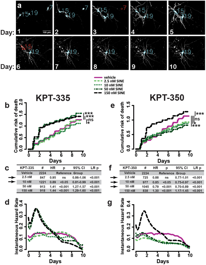
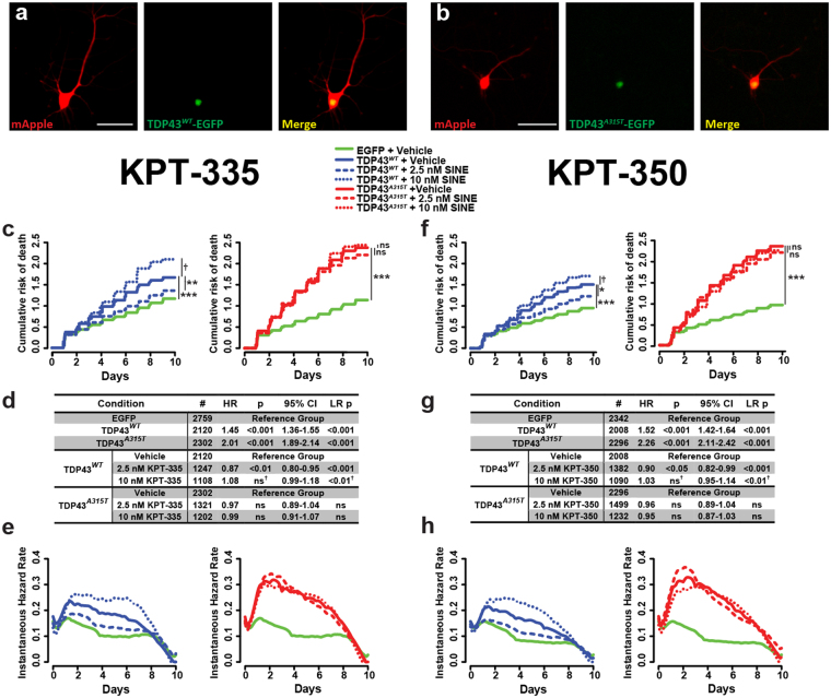
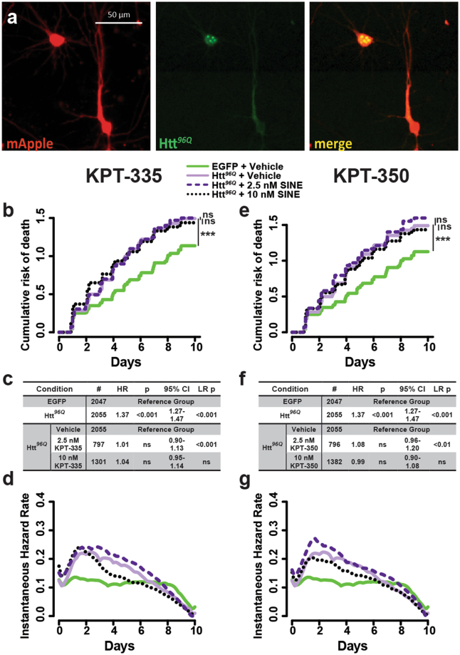
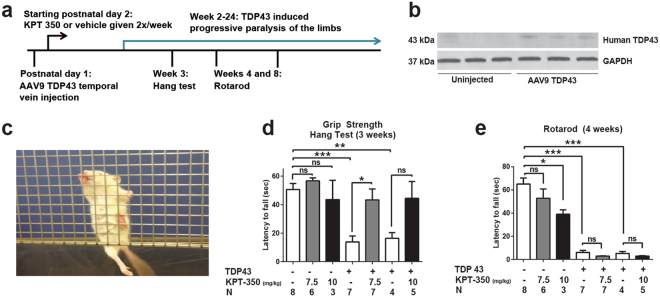
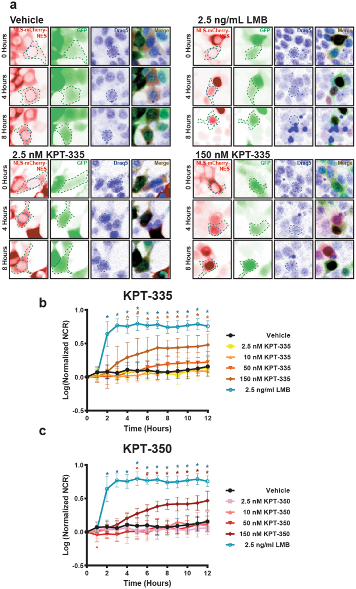
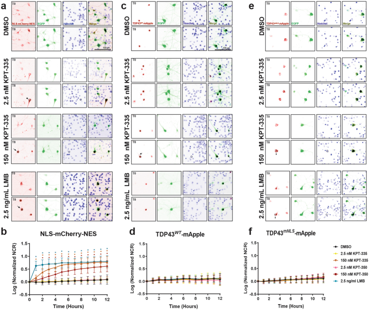
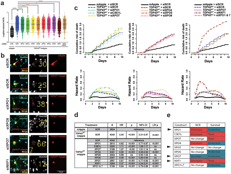
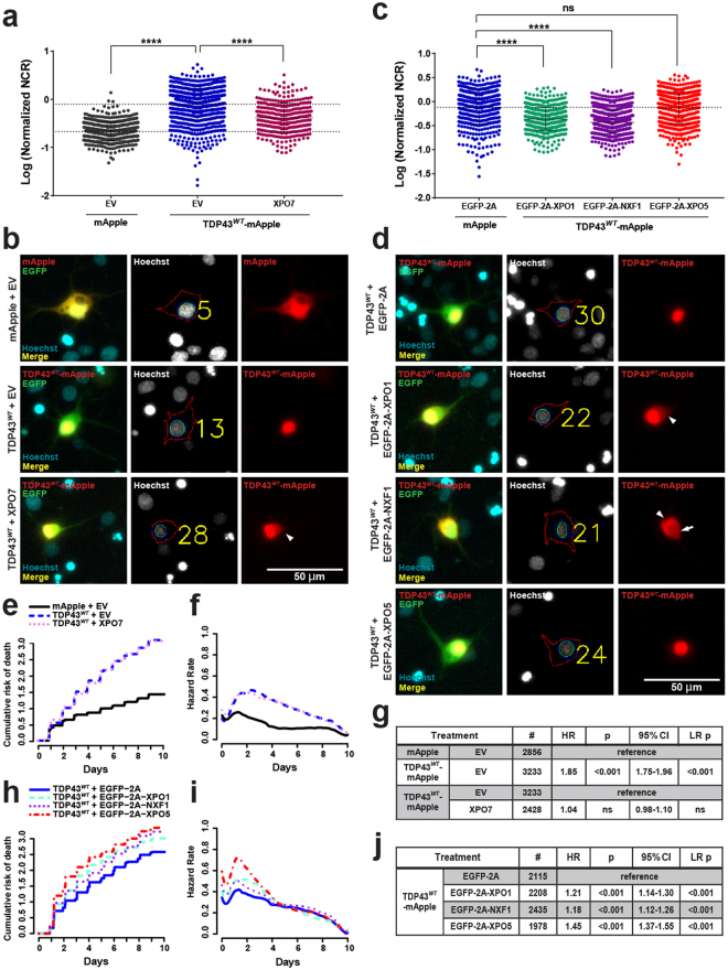
Amyotrophic lateral sclerosis (ALS) and frontotemporal dementia (FTD) are progressive neurodegenerative disorders marked in most cases by the nuclear exclusion and cytoplasmic deposition of the RNA binding protein TDP43. We previously demonstrated that ALS-associated mutant TDP43 accumulates within the cytoplasm, and that TDP43 mislocalization predicts neurodegeneration. Here, we sought to prevent neurodegeneration in ALS/FTD models using selective inhibitor of nuclear export (SINE) compounds that target exportin-1 (XPO1). SINE compounds modestly extend cellular survival in neuronal ALS/FTD models and mitigate motor symptoms in an in vivo rat ALS model. At high doses, SINE compounds block nuclear egress of an XPO1 cargo reporter, but not at lower concentrations that were associated with neuroprotection. Neither SINE compounds nor leptomycin B, a separate XPO1 inhibitor, enhanced nuclear TDP43 levels, while depletion of XPO1 or other exportins had little effect on TDP43 localization, suggesting that no single exporter is necessary for TDP43 export. Supporting this hypothesis, we find overexpression of XPO1, XPO7 and NXF1 are each sufficient to promote nuclear TDP43 egress. Taken together, our results indicate that redundant pathways regulate TDP43 nuclear export, and that therapeutic prevention of cytoplasmic TDP43 accumulation in ALS/FTD may be enhanced by targeting several overlapping mechanisms.


Conflict of interest statement

S.T. is an employee of Karyopharm Therapeutics, and has financial interest in this company. The remaining authors have no competing interests to declare.

Figures

Figure 1
Figure 1
Safety profiling of SINE compounds in rodent primary cortical neurons. (a) Primary rodent cortical neurons were transfected with EGFP and imaged every 24 h for 10 days. SINE compounds or vehicle (DMSO) were added after imaging on day 1. Neurons were identified based on morphology (blue outlines) and assigned a unique identifier (blue #). Death (red) is determined by loss of fluorescent intensity (neuron 7, day 3) or changes in morphology (neuron 15, day 6), and time of death for each cell is used to estimate the cumulative (b,e) and instantaneous (d,g) risk of death functions. Tables showing neuron number (#), hazard ratio (HR), Cox proportional hazards p value (p), confidence interval (95% CI), and log-rank p value (LR p) are shown in (c) and (f). Doses considered “safe” are indicated with black arrows. Each condition represents 3-6 combined experiments, minimum 8 technical replicates per experiment.
Figure 2
Figure 2
SINE compounds exhibit modest protective effects in models of ALS and FTD. Rodent primary cortical neurons were transfected with TDP43WT-EGFP (a), or the disease-associated mutant TDP43A315T-EGFP (b). Both TDP43WT-EGFP (solid blue line) and TDP43A315T-EGFP (solid red line) significantly increased the cumulative (c,f) and instantaneous (e,h) risk of death, compared to control neurons expressing EGFP alone (solid green line). Addition of 2.5 nM KPT-335 or KPT-350 (dashed lines) protected against toxicity from TDP43WT-EGFP (ch, blue) but not TDP43A315T-EGFP (ch, red). 10 nM SINE compound (dotted lines) enhanced the risk of death in neurons expressing TDP43WT-EGFP but not TDP43A315T-EGFP (LR test d,g). Neuron number (#), hazard ratio (HR), Cox proportional hazards p value (p), confidence interval (95% CI), and log rank p value (LR p) for each comparison are listed in (d) and (g). Each condition represents 3 combined experiments, minimum 8 technical replicates per experiment.
Figure 3
Figure 3
SINE compounds fail to protect against neurodegeneration in a model of Huntington’s disease. (a) Rodent primary cortical neurons expressing Htt96Q-EGFP, a fragment of mutant huntingtin including an expanded 96-residue polyglutamine stretch and fused to EGFP, were imaged by fluorescence microscopy. Following automated survival analysis, cumulative (b,e) and instantaneous risk of death (d,g) were determined for Htt96Q-expressing neurons (purple lines) in comparison to control cells expressing EGFP (green lines). Additional information, including the number of neurons (#), hazard ratio (HR), Cox proportional hazards p value (p), confidence interval (95% CI), and log rank p value (LR p) are shown for all experiments in (c,f). Each condition represents 3 combined experiments, 8 wells per condition, per experiment.
Figure 4
Figure 4
SINE compounds partially rescue motor deficits in an animal model of ALS/FTD. (a) Schematic timeline for in vivo experiments. (b) Western blot demonstrating the expression of human TDP43 within the brains of AAV9-injected animals, which we have previously shown is largely restricted to neuronal cell types (Jackson et al.). Full-length blots appear in Fig. S1. (c) The hang test measures the time it takes for animals to fall from grid-like scaffold when it is lifted and tilted. A single representative animal administered AAV9-TDP43 is shown; note the inability of the hind limbs to clasp the wire mesh. (d) TDP43 expression impaired motor performance on the hang test at 3 weeks, an effect that could be mitigated by 7.5 mg kg-1 KPT-350. (e) Rotarod testing at 4 weeks of age showed prominent deficits associated with TDP43 expression that were resistant to treatment with SINE compounds. n, number of animals per condition. ns, not significant, mean ± s.e.m. *p < 0.05, **p < 0.01, ***p < 0.001, one-way ANOVA with Bonferroni correction.
Figure 5
Figure 5
Nuclear localization of a reporter construct is enhanced by XPO1 inhibition. (a) The NLS-mCherry-NES fusion protein (inverted images false colored red, left panels) is localized primarily within the cytoplasm of HEK293 cells at steady-state, reflecting the dominance of the NES. Cells were cotransfected with EGFP as a cellular marker (green), and treated with the cell permeable infrared nuclear dye Draq5 (blue). Addition of 2.5 ng ml−1 leptomycin B (LMB) results in nuclear localization of the reporter by 4 h. At the 150 nM dose, KPT-355 and KPT-350 also enhanced nuclear reporter localization, albeit more slowly and less effectively than LMB. Low doses of KPT-335 and KPT-350 evoked no measurable changes in NLS-mCherry-NES localization. (a–c) The nuclear/cytoplasmic ratio (NCR) for the reporter was calculated as a function of time for HEK293 cells treated with LMB, SINE compounds or vehicle, and log transformed prior to plotting. Representative graphs in (b,c) constructed from n = 10 cells per condition, mean ± s.d., *p < 0.001, #p < 0.01, ^p < 0.05, one-way ANOVA with Dunnett’s correction.
Figure 6
Figure 6
XPO1 inhibition has no detectable effect on the subcellular distribution of TDP43. (a) Rodent primary cortical neurons were transfected with EGFP and NLS-mCherry-NES, imaged at baseline, treated with vehicle, LMB or SINE compounds, and imaged at 1 h intervals for 12 h. Representative images (false colored and inverted for clarity) are shown before (0) and 8 h after drug addition. Nuclear retention of the reporter construct is observed with LMB treatment or high (150 nM) doses of SINE compounds (black arrows, left panel at T8) but not at low (2.5 nM) doses (black arrowhead, left panel at T8). (b) LMB significantly increases the reporter nuclear to cytoplasmic ratio (NCR) by 1 h. At 150 nM, both SINE compounds significantly increase nuclear localization of NLS-mCherry-NES, with slightly slower kinetics than LMB. No change in reporter localization was observed with 2.5 nM of either SINE compound. (c,d) Following transfection with EGFP and TDP43WT-mApple, primary neurons were treated with the indicated compound and imaged by longitudinal microscopy. TDP43WT-mApple is predominantly nuclear at steady state, and SINE compounds have no observable effects on its localization. Representative image in (c) shows that no decrease in cytoplasmic TDP-43WT –mApple levels (black arrow, left panel at T8) can detected in cells treated with 150 nM KPT-335 8 h after treatment. (e,f) In primary cortical neurons, TDP43mNLS-mApple is predominantly cytoplasmic at steady-state. XPO1 inhibition fails to significantly increase TDP43mNLS-mApple NCRs at any tested dose. Nuclear clearance of TDP43mNLS-mApple was notable in treated cells, even 8 h after drug addition (black arrowheads, left panels at T8). Representative plots in b, d, f were constructed from n = 43-45 cells per condition (13-15 cells per condition, 3 replicates each), mean ± s.d., *p < 0.001, #p < 0.01, ^p < 0.05, one-way ANOVA with Dunnett’s correction.
Figure 7
Figure 7
siRNA depletion of export proteins has limited effects on TDP43 localization. (a) The distribution of TDP43WT-mApple, as estimated by the nuclear to cytoplasmic ratio (NCR) was measured 48 h after transient transfection with siRNA targeting nuclear exporters in primary rat cortical neurons. Statistical significance was calculated using 2-way ANOVA with Tukey’s adjustment. Error bars in (a) show mean ± s.d. (b) Automated analysis of subcellular protein distribution. Center panels demonstrate areas selected to measure average nuclear (within green ellipse) and cytoplasmic (between blue and red bounding lines) intensity. The effect of siRNA depletion on neuronal survival is plotted in (c). Depletion of XPO1, XPO7, or both XPO1 & 7 reduce the risk of death in transfected neurons by 20%, while NXF1 depletion more than doubles the risk of death. Cumulative hazard shown on the left, and instantaneous hazard on the right. Additional information, including the number of neurons (#), hazard ratio (HR), Cox proportional hazards p value (p), confidence interval (95% CI), and log rank p value (LR p) are shown for all experiments in (d). N = cells per condition (8-16 technical replicates per condition per experiment, minimum 3 experiments per condition). NCR was normalized to control for each biological replicate before pooling. (e) Summary of localization and survival effects. Black arrows indicate exporters chosen for further study.
Figure 8
Figure 8
XPO1, XPO7 or NXF1 overexpression enhances cytoplasmic TDP43 localization. (a,b) Overexpression of untagged XPO7 in rodent primary neurons effectively reduces the nuclear to cytoplasmic ratio (NCR) of TDP43WT-EGFP compared to cells transfected with empty vector (EV). (c,d) Overexpression of either XPO1 or NXF1 appended to the self-cleaving EGFP-2A peptide also results in significant reductions in TDP43WT-mApple NCR, while XPO5 overexpression has no effect on TDP43WT-mApple localization. Arrowheads indicate cytoplasmic TDP43 accumulation, while arrows highlight nuclear clearance of TDP43. (e–g) XPO7 overexpression has little effect on TDP43-dependent toxicity. Cumulative and instantaneous hazards shown in (e) and (f), respectively, and additional data, number of neurons (#), hazard ratio (HR), Cox proportional hazards p value (p), confidence interval (95% CI), and log rank p value (LR p) are indicated in (g). XPO1, XPO5 and NXF1 potentiate the toxicity of TDP43WT-mApple overexpression (hj). Data in a-j represent 3 biological replicates per condition, minimum 8 wells per replicate/condition. The NCR was normalized to control for each biological replicate before pooling, and plotted as mean normalized log (NCR) ± s.d. ****p < 0.0001, 2-way ANOVA with Tukey’s adjustment.

References

    1. Cima V, et al. Epidemiology of ALS in Padova district, Italy, from 1992 to 2005. Eur J Neurol. 2009;16:920–924. doi: 10.1111/j.1468-1331.2009.02623.x. - DOI - PubMed
    1. Huisman MHB, et al. Population based epidemiology of amyotrophic lateral sclerosis using capture-recapture methodology. J Neurol Neurosurg Psychiatr. 2011;82:1165–1170. doi: 10.1136/jnnp.2011.244939. - DOI - PubMed
    1. Harper CJ, Sorenson EJ, Mandrekar J. Epidemiology of amyotrophic lateral sclerosis in Minnesota: a year-long population based study. Amyotroph Lateral Scler Frontotemporal Degener. 2015;16:520–523. doi: 10.3109/21678421.2015.1051991. - DOI - PubMed
    1. Charcot JM, Joffory A. Deux cas d’atrophie musculaire progressive avec lesions de la substance grise et des faisceaux antero-lateraux de la moelle epiniere. Arch. Physiol. Neurol. Pathol. 1869;2:744–754.
    1. Lomen-Hoerth C, et al. Are amyotrophic lateral sclerosis patients cognitively normal? Neurology. 2003;60:1094–1097. doi: 10.1212/01.WNL.0000055861.95202.8D. - DOI - PubMed

Publication types

MeSH terms