. 2018 Mar 15;8(1):4641.
doi: 10.1038/s41598-018-22848-y.

Assessment of intrinsic and extrinsic signaling pathway in excitotoxic retinal ganglion cell death

Affiliations

Assessment of intrinsic and extrinsic signaling pathway in excitotoxic retinal ganglion cell death

Berkeley K Fahrenthold et al. Sci Rep. .

Abstract

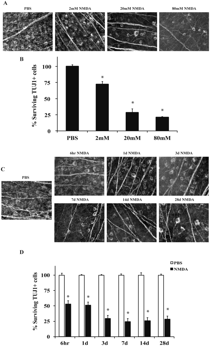
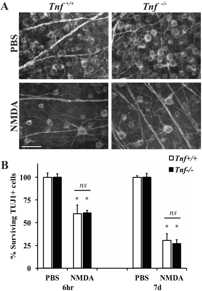
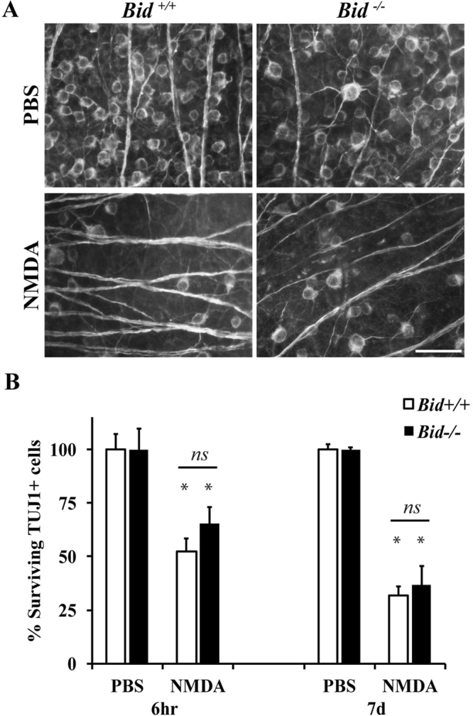
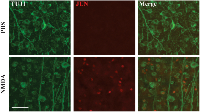
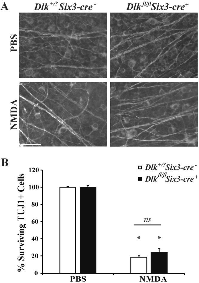
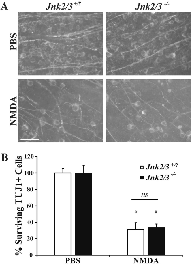
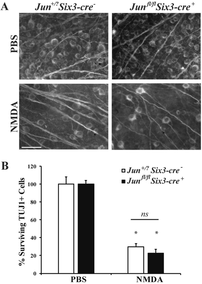
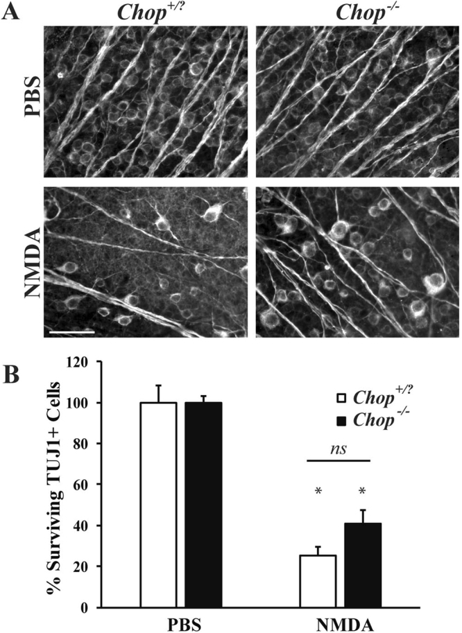
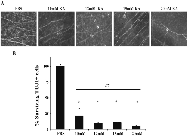
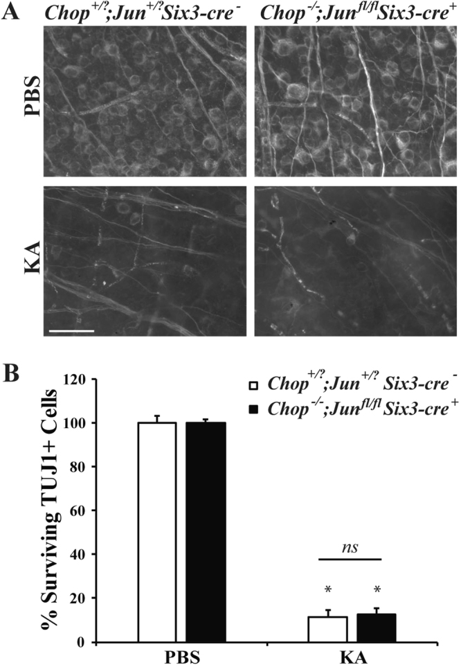
Excitotoxicity leads to the activation of a cytotoxic cascade that causes neuronal death. In the retina, retinal ganglion cells (RGCs) die after an excitotoxic insult. Multiple pathways have been proposed to contribute to RGC death after an excitotoxic insult, including TNF signaling, JNK activation, and ER stress. To test the importance of these pathways in RGC death after excitotoxic injury, the excitotoxin N-methyl-D-aspartate (NMDA) was intravitreally injected into mice deficient in components of these pathways. Absence of Tnf or its canonical downstream mediator, Bid, did not confer short- or long-term protection to RGCs. Despite known activation in RGCs and a prominent role in mediating RGC death after other insults, attenuating JNK signaling did not prevent RGC death after excitotoxic insult. Additionally, deficiency of the ER stress protein DDIT3 (CHOP), which has been shown to be involved in RGC death, did not lessen NMDA induced RGC death. Furthermore, absence of both Jun (JNK's canonical target) and Ddit3, which together provide robust, long-term protection to RGC somas after axonal insult, did not lessen RGC death. Collectively, these results indicate that the drivers of excitotoxic injury remain to be identified and/or multiple cell death pathways are activated in response to injury.


Conflict of interest statement

The authors declare no competing interests.

Figures

Figure 1
Figure 1
NMDA induced excitotoxic insult kills RGCs in a dose dependent manner. RGC loss was quantified 7 days after intravitreal injection of NMDA. (A,B) The RGC specific marker, TUJ1, was used to label RGCs in flat-mounted retinas 7 days after a 2 μl intravitreal injection of either PBS (vehicle control) or 2 mM, 20 mM, or 80 mM of NMDA into C57BL/6 J mice (n = 23 for PBS control, n = 6 for 2 mM NMDA, n = 6 for 20 mM NMDA, n = 3 for 80 mM NMDA). Quantification of TUJ1+ RGCs showed NMDA insult caused a dose dependent loss of RGCs. All NMDA concentrations caused a significant decrease in RGC number (*p < 0.001 for all comparisons). Note, that there was no significant difference in RGC number between the 20 mM and 80 mM concentrations (p = 0.819). (C,D) 20 mM NMDA was shown to induce greater than 50% RGC death at 7 days and there appeared to be no benefit to increasing the dose. Thus, 20 mM of NMDA was chosen to investigate the molecular pathways controlling NMDA induced RGC death. To understand the time course of RGC death induced by 20 mM of NMDA, TUJ1+ cells were counted in flat-mounted retinas 6hr, 1d, 3d, 7d, 14d, and 28d after (n ≥ 5 for all time points). NMDA injection resulted in significant loss of RGCs compared to controls at all time points examined (*p < 0.001). Note, approximately 50% of TUJ1 + cells were lost after 6 hours and after 3 days, no more apparent loss of TUJ1+ cells. Scale bar: 50 μm.
Figure 2
Figure 2
Tnf deficiency does not prevent RGC death after excitotoxic insult. To determine if TNF is required for NMDA induced RGC death, 2 μl PBS or 20 mM NMDA was injected into the vitreous of wildtype (Tnf+/+) and Tnf deficient (Tnf−/−) mice. The number of surviving RGCs (TUJ1 + cells) were counted 6 hours (hr) and 7 days (d) after intravitreal NMDA injection (n = 5 for 6 hr PBS Tnf+/+, n = 5 for 6 hr NMDA Tnf+/+, n = 5 for 6hr PBS Tnf−/−, n = 5 for 6hr NMDA Tnf−/−, n = 6 for 7d PBS Tnf+/+, n = 7 for 7d NMDA Tnf+/+, n = 5 for 7d PBS Tnf−/−, n = 5 for 7d NMDA Tnf−/−). (A) Representative images from flat mounted retinas stained with anti-TUJ1 show clear RGC loss in both wildtype and Tnf deficient mice after NMDA insult at both time points examined. (B) Quantification of TUJ1+ RGCs showed a significant loss of TUJ1 + cells in both wildtype and Tnf deficient mice at both 6 hours and 7 days after injury (*p < 0.001 for PBS to NMDA comparisons, P = 0.9694 comparing WT to Tnf−/− NMDA). Note, there was no difference in RGC number between Tnf wildtype and deficient mice at either time (p > 0.9 for each time point). Scale bar: 50 μm.
Figure 3
Figure 3
Bid deficiency does not prevent RGC death after excitotoxic insult. BID is a major regulator of extrinsically-induced cell death. To determine if BID is required for NMDA induced RGC death, 2 μl PBS or 20 mM NMDA was intravitreally injected into wildtype Bid (Bid+/+) and Bid deficient (Bid−/−) mice. The number of surviving RGCs (TUJ1 + cells) were counted 6 hours (hr) and 7 days (d) after intravitreal NMDA injection (n = 5 for 6 hr PBS Bid+/+, n = 5 for 6 hr NMDA Bid+/+, n = 5 for 6 hr PBS Bid−/−, n = 5 for 6 hr NMDA Bid−/−, n = 6 for 7d PBS Bid+/+, n = 6 for 7d NMDA Bid+/+, n = 7 for 7d PBS Bid−/−, n = 7 for 7d NMDA Bid−/−). (A) Representative images from flat mounted retinas stained with anti-TUJ1 show clear RGC loss in both wildtype and Bid deficient mice after NMDA insult at both time points examined. (B) Quantification of TUJ1 + RGCs showed a significant loss of TUJ1+ cells in both wildtype and Bid deficient mice at both 6 hours and 7 days after injury (*p < 0.001 for PBS to NMDA comparisons). Note, there was no difference in RGC number between Bid wildtype and deficient mice at either time (p ≥ 0.719 for each time point). Scale bar: 50 μm.
Figure 4
Figure 4
JUN signaling is activated in retinal cells in vivo following intravitreal injection of NMDA. Excitotoxic injury causes JUN activation as represented by pJUN in retinal cells, specifically RGCs, 6 hours after intravitreal injections of either NMDA. Representative images of retinal flat mounts stained with TUJ1 and JUN 6hrs after NMDA injection, showing activation of JUN in TUJ1 + cells (n = 3 for each condition). Scale bar, 50 μm.
Figure 5
Figure 5
Dlk deficiency does not protect RGCs from NMDA-induced death. (A) Representative images showing TUJ1 immunolabeled cells in the GCL of control (Dlk+/+; Six3-cre or Dlk+/fl; Six3-cre) Dlk deficient (Dlkfl/fl; Six3-cre+) mice 7 days after intravitreal NMDA or PBS injection (n = 4 for PBS WT; n = 3 for NMDA WT, n = 5 for PBS Dlkfl/fl; Six3-cre+, n = 4 for NMDA Dlkfl/fl; Six3-cre+). There was significant RGC loss in both control and Dlk deficient mice after NMDA insult. (B) Quantification of TUJ1 + RGCs showed that Dlk deficient mice had a similar loss of RGCs compared to wildtype mice 7d after NMDA injection (*p < 0.001 for comparison of PBS to NMDA; p ≥ 0.1843 for comparison between genotypes, ns between genotypes). Scale bar: 50 μm.
Figure 6
Figure 6
Jnk2−/−Jnk3−/− nulls do not have attenuated RGC loss after excitotoxic injury. (A) Representative images showing TUJ1 immunolabeled cells in the GCL of Jnk2Jnk3 deficient mice 7 days after intravitreal NMDA or PBS injection, revealing significant RGC loss in both wildtype (Jnk2+/?Jnk3+/?) and Jnk2−/−Jnk3−/− deficient mice after NMDA insult (n = 4 for PBS WT, n = 4 for NMDA WT, n = 3 for PBS Jnk2−/−Jnk3−/−, n = 3 for NMDA Jnk2−/−Jnk3−/−). (B) Quantification of TUJ1 + RGCs confirmed that Jnk2−/−Jnk3−/− deficient mice had a similar loss of RGCs compared to wildtype mice 7d after NMDA injection (*p ≤ 0.001 for comparison between PBS and NMDA; p ≥ 0.9769 comparing WT to Jnk2−/−Jnk3−/− NMDA; ns between genotypes; two-way ANOVA, Tukey’s multiple comparisons test). Scale bar: 50 μm.
Figure 7
Figure 7
Deficiency of JUN does not protect RGCs after excitotoxic injury. To determine if JUN is required for NMDA induced RGC death, NMDA was injected into the vitreous of Jun deficient (Junfl/fl; Six3-cre+) mice. The number of surviving RGCs (TUJ1+ cells) were counted 7 days after intravitreal NMDA injection. (A) Representative images from flat mounted retinas stained with anti-TUJ1 show significant RGC loss in both wildtype (Jun+/?; Six3-cre) and Junfl/fl; Six3-cre+ mice after NMDA insult (n = 5 for PBS WT, n = 5 for NMDA WT, n = 6 for PBS Junfl/fl; Six3-cre+, n = 5 for NMDA Junfl/fl; Six3-cre+). (B) Quantification of TUJ1+ RGCs confirmed that Jun deficient mice had a similar loss of RGCs compared to wildtype mice 7d after NMDA injection (*p < 0.001, comparing 20 mM NMDA to PBS control; p = 0.9846 comparing WT to Junfl/fl; Six3-cre NMDA; ns between genotypes). Scale bar: 50 μm.
Figure 8
Figure 8
Ddit3 deficiency provides modest protection of RGCs after NMDA injury. (A) Representative images showing TUJ1 immunolabeled cells in the GCL of Ddit3 deficient (Ddit3−/−), mice 7 days after intravitreal NMDA or PBS injection. Significant RGC loss occurred in wildtype (Ddit3+/?), but Ddit3 deficient mice exhibited modest protection of RGCs after NMDA insult (n = 3 for PBS WT, n = 3 for NMDA WT, n = 8 for PBS Ddit3−/−, n = 10 for NMDA Ddit3−/−). (B) Quantification of TUJ1+ RGCs confirmed that Ddit3 deficient mice had significantly more surviving RGCs compared to wildtype mice 7d after NMDA injection (*p < 0.001 comparing PBS to NMDA; p = 0.8105 comparing WT to Ddit3−/− NMDA; ns between genotypes). Scale bar: 50 μm.
Figure 9
Figure 9
Combined Jun and Ddit3 deficiency does not attenuate loss of RGCs after NMDA insult. (A) Representative images showing TUJ1 immunolabeled cells in the GCL of Jun and Ddit3 deficient (Junfl/flDdit3−/−; Six3-cre+) mice 7 days after intravitreal NMDA or PBS injection. Significant RGC loss occurred in wildtype (Jun+/?Ddit3+/?; Six3-cre) as well as Jun and Ddit3 deficient mice after NMDA insult (n = 3 for PBS WT, n = 3 for NMDA WT, n = 4 for PBS Jun+/?Ddit3+/?; Six3-cre, n = 5 for NMDA Jun+/?Ddit3+/?; Six3-cre). (B) Quantification of TUJ1+ RGCs confirmed that Jun and Ddit3 deficient mice had a similar loss of RGCs compared to wildtype mice 7d after NMDA injection (*p < 0.001 comparing PBS to NMDA; p = 0.8582 between WT and Junfl/flDdit3−/−; Six3-cre+ NMDA, ns between genotypes). Scale bar: 50 μm.
Figure 10
Figure 10
KA induced excitotoxic injury kills RGCs in a dose independent manner. RGC loss was quantified 7 days after intravitreal injection of KA. (A,B) The RGC specific marker, TUJ1, was used to label RGCs in flat-mounted retinas 7 days after a 2 μl intravitreal injection of either PBS (vehicle control) or 10 mM, 12 mM, 15, or 20 mM of KA into C57BL/6 J mice (n = 14 for PBS control, n = 6 for 10 mM KA, n = 4 for 12 mM KA, n = 5 for 15 mM KA, n = 4 for 20 mM KA). Quantification of TUJ1 + RGCs showed that KA insult caused a dose independent loss of RGCs. All KA concentrations caused a significant decrease in RGC number (*p < 0.001 for all concentrations compared to control). Scale bar: 50 μm.
Figure 11
Figure 11
Combined Jun and Ddit3 deficiency does not attenuate loss of RGCs after KA insult. (A) Representative images showing TUJ1 immunolabeled cells in the GCL of Jun and Ddit3 deficient (Junfl/flDdit3−/−; Six3-cre+) mice 7 days after intravitreal KA or PBS injection. Significant RGC loss occurred in wildtype (Jun+/?Ddit3+/?; Six3-cre) as well as Jun and Ddit3 deficient mice after KA insult (n = 5 for PBS WT, n = 3 for NMDA WT, n = 4 for PBS Jun+/?Ddit3+/?; Six3-cre, n = 3 for NMDA Jun+/?Ddit3+/?; Six3-cre). (B) Quantification of TUJ1+ RGCs confirmed that Jun and Ddit3 deficient mice had a similar loss of RGCs compared to wildtype mice 7d after NMDA injection (*p < 0.001 comparing PBS to KA; p = 0.9962 between WT and Junfl/flDdit3−/−; Six3-cre+ KA, ns between genotypes). Scale bar: 50 μm.

Similar articles

Cited by

References

    1. Olney JW. Brain lesions, obesity, and other disturbances in mice treated with monosodium glutamate. Science. 1969;164:719–721. doi: 10.1126/science.164.3880.719. - DOI - PubMed
    1. Araszkiewicz, A. & Zozulinska-Ziolkiewicz, D. Retinal neurodegeneration in the course of diabetes - pathogenesis and clinical perspective. Curr. Neuropharmacol. (2016). - PMC - PubMed
    1. Barber AJ. A new view of diabetic retinopathy: a neurodegenerative disease of the eye. Prog. Neuropsychopharmacol. Biol. Psychiatry. 2003;27:283–290. doi: 10.1016/S0278-5846(03)00023-X. - DOI - PubMed
    1. Ishikawa M. Abnormalities in Glutamate Metabolism and Excitotoxicity in the Retinal Diseases. Scientifica. 2013;2013:e528940. doi: 10.1155/2013/528940. - DOI - PMC - PubMed
    1. Fischer AJ, Pickett Seltner RL, Poon J, Stell WK. Immunocytochemical characterization of quisqualic acid- and N-methyl-D-aspartate-induced excitotoxicity in the retina of chicks. J. Comp. Neurol. 1998;393:1–15. doi: 10.1002/(SICI)1096-9861(19980330)393:1<1::AID-CNE1>3.0.CO;2-3. - DOI - PubMed

Publication types

Substances