

Monetary Reward and Punishment to Response Inhibition Modulate Activation and Synchronization Within the Inhibitory Brain Network
- PMID: 29545745
- PMCID: PMC5837970
- DOI: 10.3389/fnhum.2018.00027
Monetary Reward and Punishment to Response Inhibition Modulate Activation and Synchronization Within the Inhibitory Brain Network
Abstract
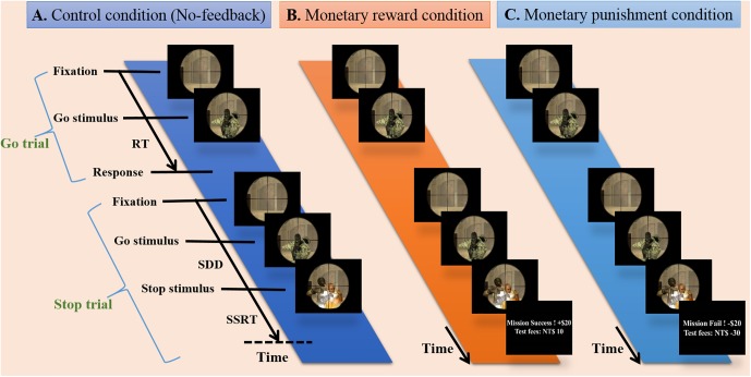
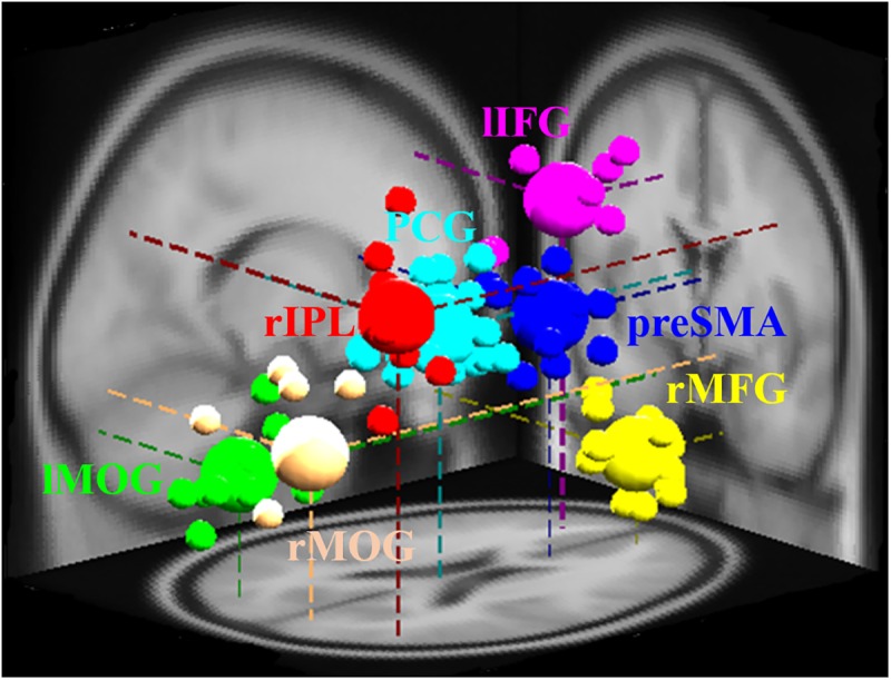
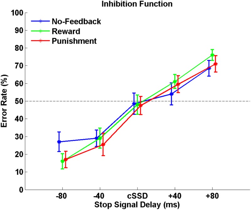
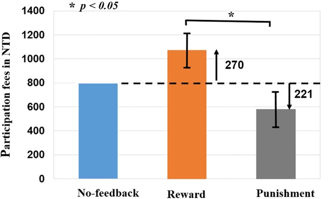
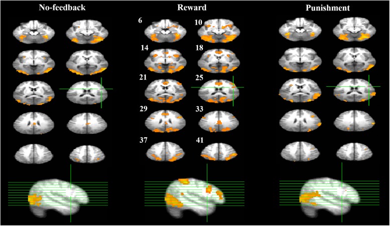
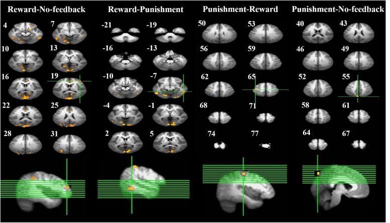
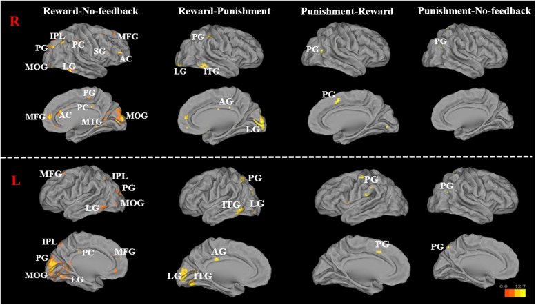
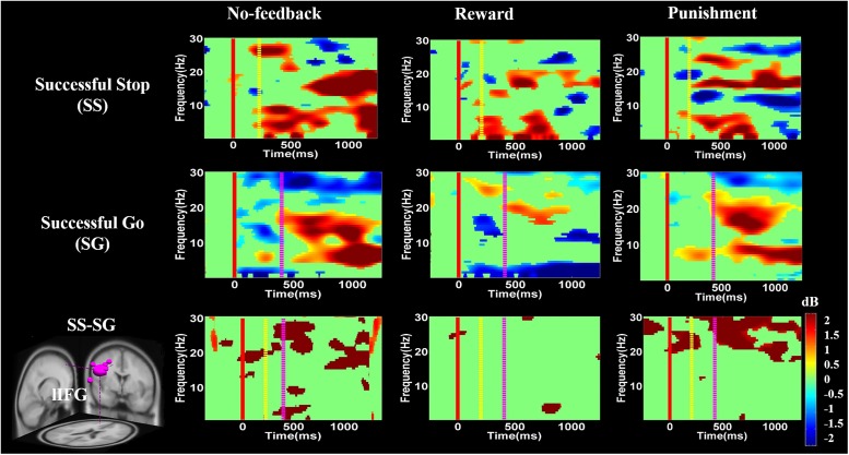
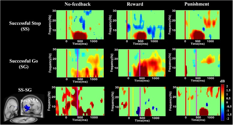
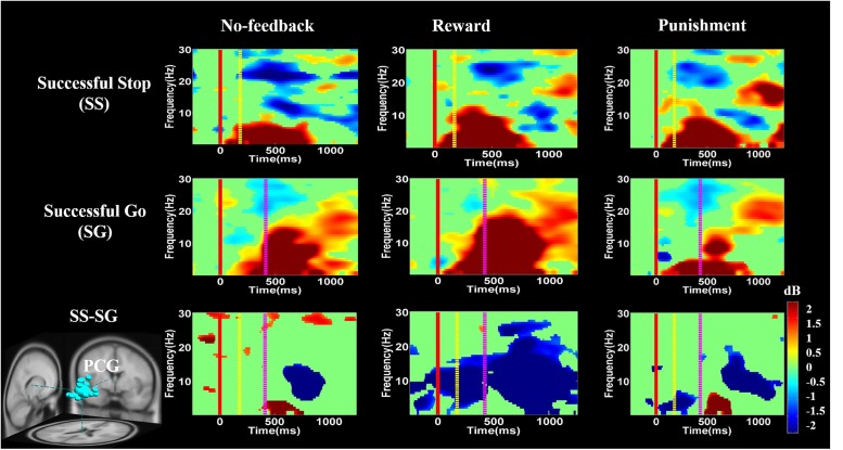
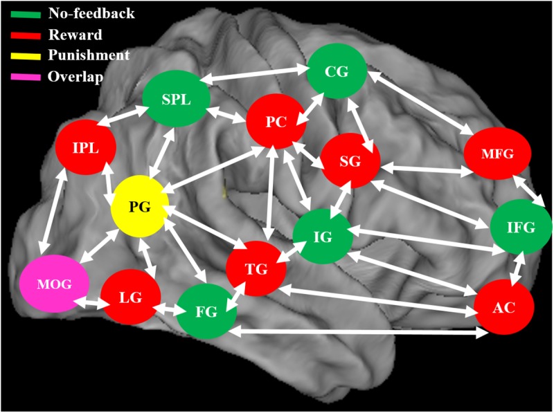
A reward or punishment can modulate motivation and emotions, which in turn affect cognitive processing. The present simultaneous functional magnetic resonance imaging-electroencephalography study examines neural mechanisms of response inhibition under the influence of a monetary reward or punishment by implementing a modified stop-signal task in a virtual battlefield scenario. The participants were instructed to play as snipers who open fire at a terrorist target but withhold shooting in the presence of a hostage. The participants performed the task under three different feedback conditions in counterbalanced order: a reward condition where each successfully withheld response added a bonus (i.e., positive feedback) to the startup credit, a punishment condition where each failure in stopping deduced a penalty (i.e., negative feedback), and a no-feedback condition where response outcome had no consequences and served as a control setting. Behaviorally both reward and punishment conditions led to significantly down-regulated inhibitory function in terms of the critical stop-signal delay. As for the neuroimaging results, increased activities were found for the no-feedback condition in regions previously reported to be associated with response inhibition, including the right inferior frontal gyrus and the pre-supplementary motor area. Moreover, higher activation of the lingual gyrus, posterior cingulate gyrus (PCG) and inferior parietal lobule were found in the reward condition, while stronger activation of the precuneus gyrus was found in the punishment condition. The positive feedback was also associated with stronger changes of delta, theta, and alpha synchronization in the PCG than were the negative or no-feedback conditions. These findings depicted the intertwining relationship between response inhibition and motivation networks.
Keywords: electroencephalography; functional magnetic resonance imaging; motivation; no-feedback; posterior cingulate gyrus; punishment; response inhibition; reward.
Figures
Similar articles
-
Effects of reward and punishment on brain activations associated with inhibitory control in cigarette smokers.Addiction. 2013 Nov;108(11):1969-78. doi: 10.1111/add.12276. Epub 2013 Jul 12. Addiction. 2013. PMID: 23773427
-
Mapping social reward and punishment processing in the human brain: A voxel-based meta-analysis of neuroimaging findings using the social incentive delay task.Neurosci Biobehav Rev. 2021 Mar;122:1-17. doi: 10.1016/j.neubiorev.2020.12.034. Epub 2021 Jan 6. Neurosci Biobehav Rev. 2021. PMID: 33421544 Review.
-
Event-related components of the punishment and reward sensitivity.Clin Neurophysiol. 2010 Jan;121(1):60-76. doi: 10.1016/j.clinph.2009.10.004. Epub 2009 Nov 8. Clin Neurophysiol. 2010. PMID: 19900840
-
Alterations of monetary reward and punishment processing in chronic cannabis users: an FMRI study.PLoS One. 2015 Mar 23;10(3):e0119150. doi: 10.1371/journal.pone.0119150. eCollection 2015. PLoS One. 2015. PMID: 25799565 Free PMC article.
-
Reward positivity: Reward prediction error or salience prediction error?Psychophysiology. 2016 Aug;53(8):1185-92. doi: 10.1111/psyp.12673. Epub 2016 May 17. Psychophysiology. 2016. PMID: 27184070 Review.
Cited by
-
Early Imaging Based Predictive Modeling of Cognitive Performance Following Therapy for Childhood ALL.IEEE Access. 2019;7:146662-146674. doi: 10.1109/access.2019.2946240. Epub 2019 Oct 8. IEEE Access. 2019. PMID: 32547892 Free PMC article.
-
Alzheimer's Disease Analysis Algorithm Based on No-threshold Recurrence Plot Convolution Network.Front Aging Neurosci. 2022 May 10;14:888577. doi: 10.3389/fnagi.2022.888577. eCollection 2022. Front Aging Neurosci. 2022. PMID: 35619941 Free PMC article.
-
Neural Dynamics of Target Detection via Wireless EEG in Embodied Cognition.Sensors (Basel). 2021 Jul 31;21(15):5213. doi: 10.3390/s21155213. Sensors (Basel). 2021. PMID: 34372448 Free PMC article.
-
The effect of task complexity on the neural network for response inhibition: An ALE meta-analysis.Neurosci Biobehav Rev. 2024 Mar;158:105544. doi: 10.1016/j.neubiorev.2024.105544. Epub 2024 Jan 12. Neurosci Biobehav Rev. 2024. PMID: 38220034 Free PMC article. Review.
-
Noisy Galvanic Vestibular Stimulation (Stochastic Resonance) Changes Electroencephalography Activities and Postural Control in Patients with Bilateral Vestibular Hypofunction.Brain Sci. 2020 Oct 15;10(10):740. doi: 10.3390/brainsci10100740. Brain Sci. 2020. PMID: 33076417 Free PMC article.
References
LinkOut - more resources
Full Text Sources
Other Literature Sources
Research Materials