. 2018 Mar 1:9:387.
doi: 10.3389/fimmu.2018.00387. eCollection 2018.

Systemic Lupus Erythematosus: Definitions, Contexts, Conflicts, Enigmas

Affiliations

Systemic Lupus Erythematosus: Definitions, Contexts, Conflicts, Enigmas

Ole Petter Rekvig. Front Immunol. .

Abstract

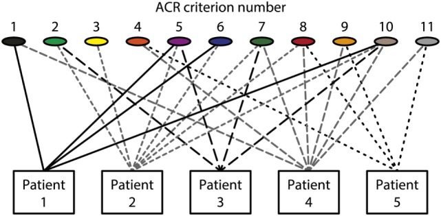
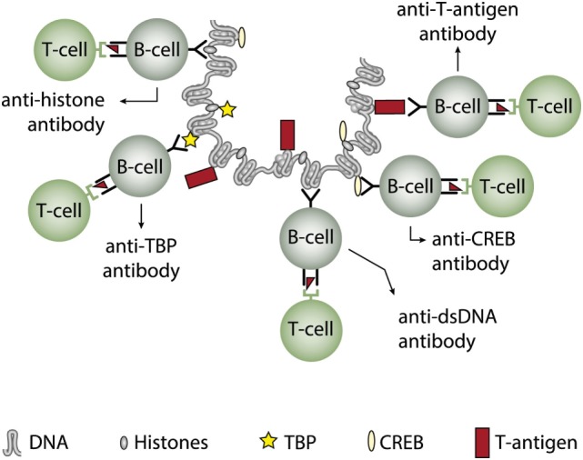
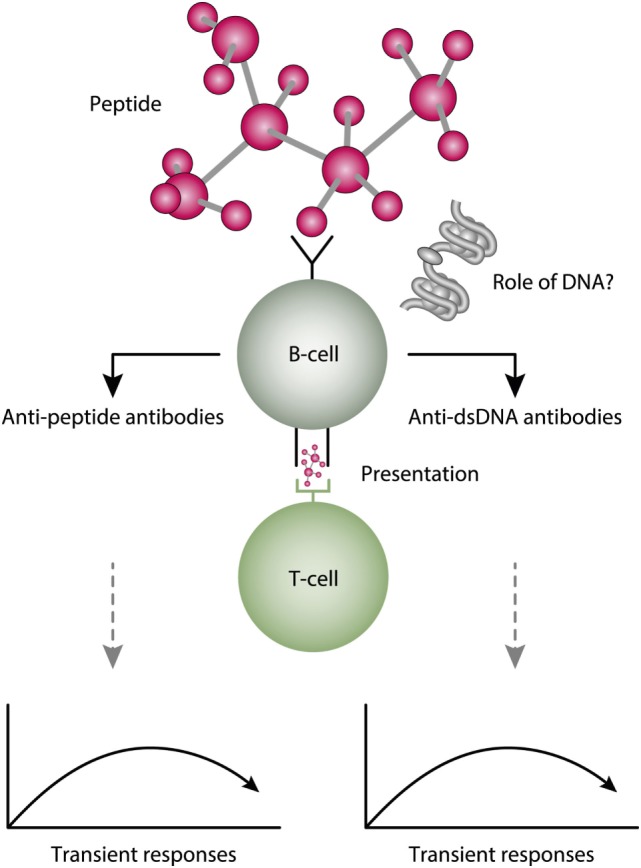
Systemic lupus erythematosus (SLE) is an inadequately defined syndrome. Etiology and pathogenesis remain largely unknown. SLE is on the other hand a seminal syndrome that has challenged immunologists, biologists, genetics, and clinicians to solve its nature. The syndrome is characterized by multiple, etiologically unlinked manifestations. Unexpectedly, they seem to occur in different stochastically linked clusters, although single gene defects may promote a smaller spectrum of symptoms/criteria typical for SLE. There is no known inner coherence of parameters (criteria) making up the disease. These parameters are, nevertheless, implemented in The American College of Rheumatology (ACR) and The Systemic Lupus Collaborating Clinics (SLICC) criteria to classify SLE. Still, SLE is an abstraction since the ACR or SLICC criteria allow us to define hundreds of different clinical SLE phenotypes. This is a major point of the present discussion and uses "The anti-dsDNA antibody" as an example related to the problematic search for biomarkers for SLE. The following discussion will show how problematic this is: the disease is defined through non-coherent classification criteria, its complexity is recognized and accepted, its pathogenesis is plural and poorly understood. Therapy is focused on dominant symptoms or organ manifestations, and not on the syndrome itself. From basic scientific evidences, we can add substantial amount of data that are not sufficiently considered in clinical medicine, which may change the paradigms linked to what "The Anti-DNA antibody" is-and is not-in context of the imperfectly defined syndrome SLE.

Keywords: anti-dsDNA antibodies; criteria; definitions; enigma; syndrome; systemic lupus erythematosus.


Figures

Figure 1
Figure 1
Patients classified to have systemic lupus erythematosus (SLE) by the The American College of Rheumatology (ACR) classification criteria—diversity of the clinical phenotypes. On top of the figure, each of the 11 ACR criteria is presented symbolically (see Table 1 for details on the ACR criteria). Five patients are demonstrated. The patients share some criteria, but diverge with respect to others. This chaotic figure demonstrates that the use of criteria is dubious to investigate pathogenesis of the syndrome and to search for biomarkers to characterize the syndrome SLE. How can we determine common features or biomarkers, when SLE presents so many different phenotypes? The patients in this figure are fictive but they reflect problems with the ACR in real life.
Figure 2
Figure 2
Theoretical anti-dsDNA antibody profiles in context of systemic lupus erythematosus (SLE) classification criteria. The American College of Rheumatology (ACR) and SLICC SLE classification criteria include anti-dsDNA antibodies as a criterium. As a criterium the antibodies are poorly defined. For example, a short-lived stimulus by an infectious agent may induce transient antibodies at low titers (A). If the infectious stimulus prevails, the anti-dsDNA antibody may prevail at low titers, even though above the assay cutoff level (B). The anti-dsDNA antibody production in (C) is transient, although at high titers, as a consequence of a strong, transient stimulus either of autologous or, e.g., infectious origin. In (D), the immune response is characterized by sustained production of anti-dsDNA antibodies at high to very high titers. The red parts of each profile represent autoantibody levels above the antibody cut-off levels as defined by ACR or SLICC criteria. The curves are fictive and constructed empirically in order to demonstrate the variability of anti-dsDNA antibody profiles, all of which fulfill requirements in the ACR and SLICC classification criteria for SLE. See text for details.
Figure 3
Figure 3
Cognate interaction of DNA-specific B cells and bacterial-derived peptide-specific T cells. This example describes a classical hapten-carrier-like model to explain production of anti-dsDNA antibodies in non-SLE (left panel) and in SLE conditions (right panel). In this model, chromatin-associated dsDNA functions as a non-immunogenic hapten that is recognized by the B cell antigen receptor, while heterologous, bacterial DNA-binding protein-derived peptides function as carrier proteins that activate peptide-specific T helper cells. In this scenario, T cell tolerance for nucleosomes is maintained intact, and the immune response is transient and is limited to the duration of the bacterial infection. According to Pisetsky et al. (83, 135), the immune response is dichotomous in the sense that in a normal immunogenic context, the antibodies recognize bacterial DNA (left part of the panel), while in an autoimmune (SLE-like) context antibodies are also produced that recognize mammalian dsDNA (right part of the panel). These processes may be operational in vivo in experimental and native contexts.
Figure 4
Figure 4
Cognate interaction of DNA-specific B cells and DNA-binding and virus-derived peptide-specific T cells. The figure presents two variants of a classical hapten-carrier-like model. In this model, chromatin from virus-infected cells are released in complex with DNA-binding viral proteins (left panel). Cognate interaction of mammalian dsDNA-specific B cells and virus peptide-specific T cells result in production of antimammalian dsDNA-specific antibodies. This process may be operational in genetically normal individuals (115, 117). In the right panel, the virus-infected cells also release viral mini chromosomes in complex with viral proteins. B cells recognize viral DNA, and present processed DNA-bound virus-encoded peptides to non-tolerant T cells. In this situation, antivirus DNA antibodies are produced. The antibody profiles depend on the time-line and kinetics of the virus infection. From this, lupus-like anti-dsDNA antibodies may appear in non-lupus individuals, thus questioning the validity of pure detection of anti-dsDNA antibodies as a SLE classification criterion.
Figure 5
Figure 5
Induction of anti-dsDNA antibodies by in vivo expression of a single viral dsDNA-binding protein. Injection of normal mice with plasmids encoding wild type polyomavirus DNA-binding T antigen in context of eukaryotic promoters induced production of antibodies to T antigen and significant production of antibodies to mammalian dsDNA, histones, and to certain transcription factors like TATA-binding protein (TBP) and cAMP-responsive element-binding protein (CREB). All autologous chromatin-derived ligands physically linked to T antigen can therefore be rendered immunogenic to autoimmune B cells that present peptides derived from T antigen. Therefore, concerted production of autoantibodies specific for chromatin antigens, including dsDNA and histones, is not depending on a systemic lupus erythematosus background, but may appear also in quite healthy individuals.
Figure 6
Figure 6
A theoretical model to explain peptide-induced anti-dsDNA and anti-peptide antibodies. Some peptides have the property to act as inducers of anti-dsDNA antibodies. There are problems with this cross-stimulating model, since it is not obvious that the peptide-induced immune response will affinity maturate toward dsDNA. Rather, somatic hypermutations in the variable heavy chain complementary determining regions (VH CDR) may shift the dual specificity toward a focused specificity for the peptide. Whether chromatin (indicated in the figure) may drive the peptide-induced anti-dsDNA antibody further is unlikely from two reasons. For the first, the initial response is controlled by peptide-specific, and not by chromatin-specific T cells. Second, if chromatin was not involved in early phases of the responses, it is no reason to believe it is rendered immunogenic in later phases of the responses.

References

    1. Rekvig OP, Nossent JC. Anti-double-stranded DNA antibodies, nucleosomes, and systemic lupus erythematosus: a time for new paradigms? Arthritis Rheum (2003) 48:300–12.10.1002/art.10739 - DOI - PubMed
    1. Rekvig OP, van der Vlag J. The pathogenesis and diagnosis of systemic lupus erythematosus: still not resolved. Semin Immunopathol (2014) 36:301–11.10.1007/s00281-014-0428-6 - DOI - PubMed
    1. Goilav B, Putterman C. The role of anti-DNA antibodies in the development of lupus nephritis: a complementary, or alternative, viewpoint? Semin Nephrol (2015) 35:439–43.10.1016/j.semnephrol.2015.08.005 - DOI - PMC - PubMed
    1. Marion TN, Postlethwaite AE. Chance, genetics, and the heterogeneity of disease and pathogenesis in systemic lupus erythematosus. Semin Immunopathol (2014) 36:495–517.10.1007/s00281-014-0440-x - DOI - PubMed
    1. Petri M, Orbai AM, Alarcon GS, Gordon C, Merrill JT, Fortin PR, et al. Derivation and validation of the Systemic Lupus International Collaborating Clinics classification criteria for systemic lupus erythematosus. Arthritis Rheum (2012) 64:2677–86.10.1002/art.34473 - DOI - PMC - PubMed

Publication types