. 2018 Apr;15(4):3376-3384.
doi: 10.3892/etm.2018.5815. Epub 2018 Jan 30.

Effect of TLR4/MyD88 signaling pathway on sepsis-associated acute respiratory distress syndrome in rats, via regulation of macrophage activation and inflammatory response

Affiliations

Effect of TLR4/MyD88 signaling pathway on sepsis-associated acute respiratory distress syndrome in rats, via regulation of macrophage activation and inflammatory response

Shujun Zhou et al. Exp Ther Med. 2018 Apr.

Abstract

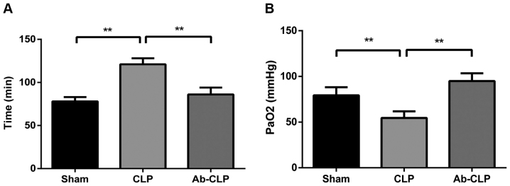
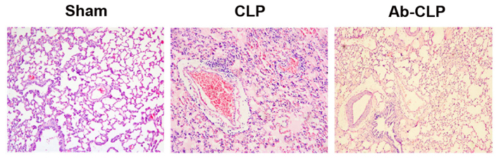
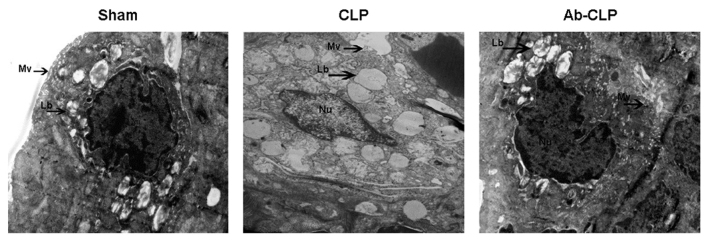
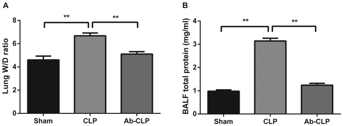
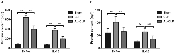
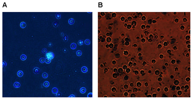
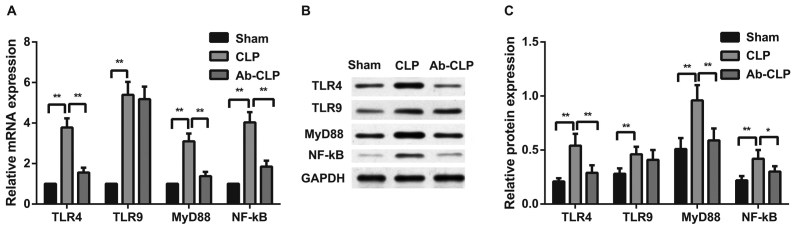
The present study aimed to investigate the effects of the Toll-like receptor (TLR)4/myeloid differentiation primary response (MyD)88 signaling pathway on sepsis-associated acute respiratory distress syndrome (ARDS) in rats, and the involvement of macrophage activation and the inflammatory response. A total of 36 specific pathogen-free male Sprague-Dawley rats were selected to establish the rat model of sepsis-associated ARDS using cecal ligation and puncture (CLP). Rats were assigned into the Ab (anti-TLR4 monoclonal antibody)-CLP, CLP and Sham groups. Arterial partial pressure of oxygen (PaO2) was detected using blood gas analysis. Bronchoalveolar lavage fluid (BALF) and alveolar macrophages were collected. The pathological structure of lung tissue was observed following hematoxylin-eosin staining. The ultrastructural alterations of alveolar epithelial cells were observed under transmission electron microscope. The ratios of wet/dry weight of lung tissue and total protein content in BALF were measured. The concentration of tumor necrosis factor (TNF)-α and interleukin (IL)-1β in BALF and peripheral blood was determined by enzyme-linked immunosorbent assay. The TLR4, TLR9, MyD88 and nuclear factor (NF)-κΒ mRNA and protein expression levels in alveolar macrophages were measured by reverse transcription-quantitative polymerase chain reaction and western blotting. Compared with the Sham group, the rats in the CLP group demonstrated significantly increased respiratory frequency, lung permeability, lung edema, inflammatory infiltration, TNF-α and IL-1β expression levels in BALF and peripheral blood and TLR4, TLR9, MyD88 and NF-κΒ expression levels in macrophages, however decreased arterial PaO2. Following pretreatment with anti-TLR4 monoclonal antibody, rats exhibited decreased lung injury, inflammatory infiltration, lung edema, TNF-α and IL-1β expressions in BALF and peripheral blood, and TLR4, TLR9, MyD88 and NF-κΒ expression levels in macrophages, with increased arterial PaO2. These results suggested that the inhibition of TLR4/MyD88 signaling pathway may relieve sepsis-associated ARDS in rats through regulating macrophage activation and the inflammatory response.

Keywords: TLR4/MyD88 signaling pathway; acute respiratory distress syndrome; inflammatory response; macrophage activation; sepsis.


Figures

Figure 1.
Figure 1.
Respiratory frequency and arterial PaO2 of rats in three groups. (A) Comparison of respiratory frequency of rats among the Sham, CLP and Ab-CLP groups; (B) comparison of the arterial PaO2 of rats among the Sham, CLP and Ab-CLP groups by blood gas analysis; **P<0.005 compared with the CLP group. PaO2, partial pressure of oxygen; CLP, cecal ligation puncture; Ab, anti-TLR4 monoclonal antibody.
Figure 2.
Figure 2.
Pathological structure of lung tissue of rats by H&E staining in three groups (magnification, ×200). H&E, hematoxylin and eosin; CLP, cecal ligation puncture; Ab, anti-TLR4 monoclonal antibody.
Figure 3.
Figure 3.
Cellular structure of type II alveolar epithelial cells of rats in three groups under transmmision electron microscope (magnification, ×2,800). CLP, cecal ligation puncture; Ab, anti-TLR4 monoclonal antibody; Lb, Lamellar bodies; Mv, Microvilli; Nu, Nucleus.
Figure 4.
Figure 4.
Comparisons of W/D ratios and total protein content in BALF of rats in three groups. (A) Comparison of the W/D ratios of rats among the Sham, CLP and Ab-CLP groups; (B) comparison of the total protein content in BALF of rats among the Sham, CLP and Ab-CLP groups; **P<0.005 compared with the CLP group. W/D, wet weight/dry weight; BALF, bronchoalveolar lavage fluid; CLP, cecal ligation puncture; Ab, anti-TLR4 monoclonal antibody.
Figure 5.
Figure 5.
Comparison of the protein contents of inflammatory factors c in BALF and peripheral blood among the three groups. (A) Comparison of the protein contents of inflammatory factors TNF-α and IL-1β in BALF among the Sham, CLP and Ab-CLP groups; (B) comparison of the protein contents of inflammatory factors TNF-α and IL-1β in peripheral blood among the Sham, CLP and Ab-CLP groups; **P<0.005 compared with the CLP group; ***P<0.001 compared with the CLP group. BALF, bronchoalveolar lavage fluid; CLP, cecal ligation puncture; Ab, anti-TLR4 monoclonal antibody; TNF-α, tumor necrosis factor-α; IL-1β, interleukin-1β.
Figure 6.
Figure 6.
Images of alveolar macrophage after wright-giemsa staining and trypan blue staining (magnification, ×400). (A) Image of alveolar macrophage after wright-giemsa staining; (B) image of alveolar macrophage after trypan blue staining.
Figure 7.
Figure 7.
Comparison of the mRNA and protein expressions of TLR4/MyD88 signaling pathway-related factors in alveolar macrophage among the three groups. (A) Comparison of TLR4, TLR9, MyD88 and NF-κΒ P65 mRNA expressions in alveolar macrophage of rats among the Sham, CLP and Ab-CLP groups; (B) protein bands of TLR4, TLR9, MyD88 and NF-κΒ P65 in the Sham, CLP and Ab-CLP detected by western blotting; (C) comparison of TLR4, TLR9, MyD88 and NF-κΒ P65 protein expressions in alveolar macrophage of rats among the Sham, CLP and Ab-CLP groups; **P<0.005 compared with the CLP group; *P<0.05 compared with the CLP group. CLP, cecal ligation puncture; Ab, anti-TLR-4 monoclonal antibody; TLR4, Toll-like receptor 4; TLR9, Toll-like receptor 9; MyD88, myeloid differentiation primary-response protein 88; NF-κB, nuclear factor-κB; GAPDH, glyceraldehyde-3-phosphate dehydrogenase.

References

    1. Han S, Mallampalli RK. The acute respiratory distress syndrome: From mechanism to translation. J Immunol. 2015;194:855–860. doi: 10.4049/jimmunol.1402513. - DOI - PMC - PubMed
    1. Lin WC, Chen CW, Huang YW, Chao L, Chao J, Lin YS, Lin CF. Kallistatin protects against sepsis-related acute lung injury via inhibiting inflammation and apoptosis. Sci Rep. 2015;5:12463. doi: 10.1038/srep12463. - DOI - PMC - PubMed
    1. Wang M, Yan J, He X, Zhong Q, Zhan C, Li S. Candidate genes and pathogenesis investigation for sepsis-related acute respiratory distress syndrome based on gene expression profile. Biol Res. 2016;49:25. doi: 10.1186/s40659-016-0085-4. - DOI - PMC - PubMed
    1. Xu Z, Huang Y, Mao P, Zhang J, Li Y. Sepsis and ARDS: The dark side of histones. Mediators Inflamm. 2015;2015:205054. doi: 10.1155/2015/205054. - DOI - PMC - PubMed
    1. Boyle AJ, Mac Sweeney R, McAuley DF. Pharmacological treatments in ARDS; a state-of-the-art update. BMC Med. 2013;11:166. doi: 10.1186/1741-7015-11-166. - DOI - PMC - PubMed