. 2018 Apr;15(4):3404-3412.
doi: 10.3892/etm.2018.5819. Epub 2018 Jan 31.

Protective effects of amifostine, curcumin and caffeic acid phenethyl ester against cisplatin-induced testis tissue damage in rats

Affiliations

Protective effects of amifostine, curcumin and caffeic acid phenethyl ester against cisplatin-induced testis tissue damage in rats

Tolga Mercantepe et al. Exp Ther Med. 2018 Apr.

Abstract

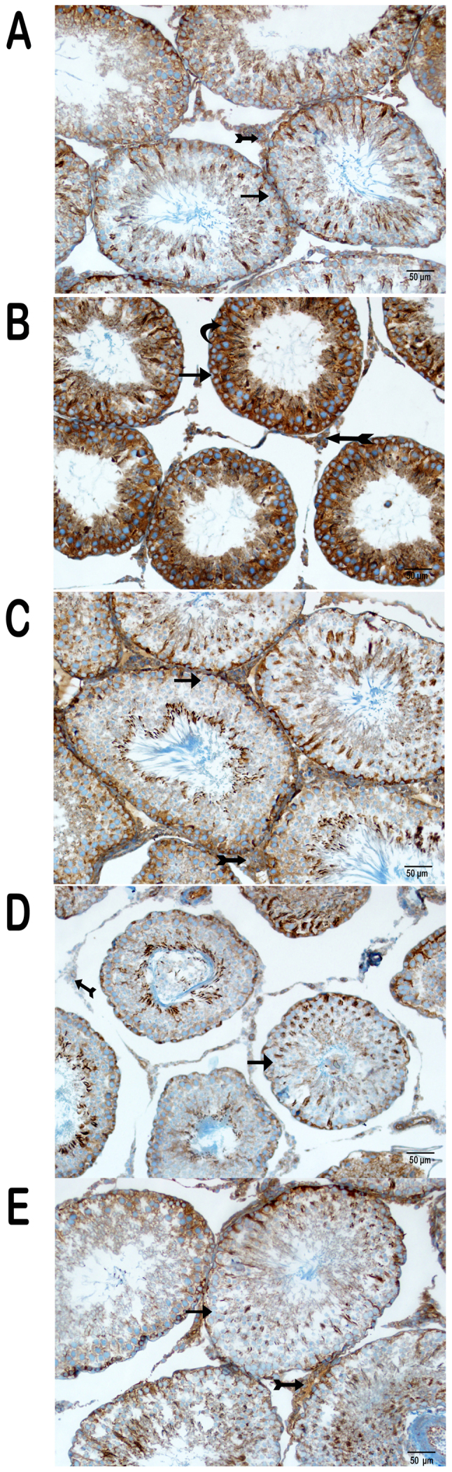
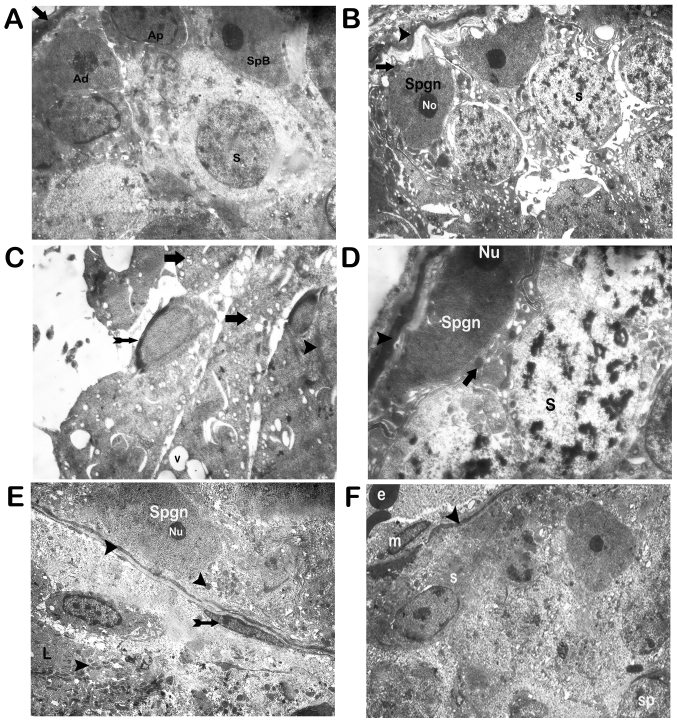
Cisplatin is an effective antineoplastic drug that is usually used to treat a number of different types of cancer in the clinic. One of the most notable side effects of cisplatin use is infertility. The present study was designed to determine the non-oxidative testicular effects caused by the use of cisplatin in rats. The rats were randomly allocated to the experimental groups. The untreated rats represented the control group (group I) and the treatment groups were as follows: cisplatin alone (group II), cisplatin+amifostine (group III), cisplatin+curcumin (group IV), and cisplatin+caffeic acid phenethyl ester (CAPE; group V). The present study observed that following cisplatin administration, the expression of nuclear factor-κB (NF-κβ)/p65, caspase-3 and 8-deoxyguanosine (8-OHdG) increased in germinal epithelium and Leydig cells. However, the expression of these markers decreased in groups III-V, most notably in the group treated with amifostine. cisplatin induced-damage was countered by amifostine and curcumin. The results revealed that the activation of NF-κB, caspase-3 and 8-OHdG had a significant role in cisplatin-induced testicular toxicity. Thus, amifostine, curcumin and, to a lesser extent, CAPE have the potential for use as therapeutic adjuvants in cisplatin-induced testis injury.

Keywords: amifostine; caffeic acid phenethyl ester; cisplatin; curcumin; testis.


Figures

Figure 1.
Figure 1.
Light microscope images of testicular tissue by hematoxylin and eosin staining. (A) Control group (magnification, ×20). (B) Control group (magnification, ×40). (C) Cis applied group: Atrophic seminiferous tubules (magnification, ×40). (D) Cis+amifostine applied group (magnification, ×40). (E) Cis+curcumin applied group (magnification, ×20). (F) Cis+CAPE applied group (magnification, ×40). Sertoli cells are indicated by spirally arrows; basal lamina of the seminiferous tubule with the myoepithelial cell are indicated by an arrow; spermatogonia are indicated by a thin arrow; spermatids are indicated by an arrowhead; Leydig cells are indicated by a tailed arrow; primary spermatocytes are indicated by curved arrows; Hyalinized interstitial areas are indicated by a star; Oedematous fields are indicated by an asterisk; Cis, cisplatin; CAPE, caffeic acid phenethyl ester.
Figure 2.
Figure 2.
Light microscope images of testicular tissue with Periodic Acid Schiff staining. (A) Control group (magnification, 40x). (B) Cis applied group: Atrophic seminiferous tubules. Irregular basal lamina of the seminiferous tubules (indicated by an arrow). Spermatogonia with condensed chromatin (indicated by a thin arrow; magnification, 20x). (C) Cis+amifostine applied group: Regular basal lamina of the seminiferous tubule (indicated by an arrow; magnification, 20x). (D) Cis+curcumin applied group (magnification, ×40). (E) Cis+CAPE applied group (magnification, 20x). Sertoli cells are indicated by spirally arrows; basal lamina of the seminiferous tubule with the myoepithelial cell are indicated by an arrow; spermatogonia are indicated by a thin arrow; spermatids are indicated by an arrowhead; Leydig cells are indicated by a tailed arrow; primary spermatocytes are indicated by curved arrows; Hyalinized interstitial areas are indicated by a star; Oedematous fields are indicated by an asterisk; Cis, cisplatin; CAPE, caffeic acid phenethyl ester.
Figure 3.
Figure 3.
Light microscope images of testicular tissue with nuclear factor-κB/p65 staining. (A) Control group (magnification, 20x). (B) Cis applied group (magnification, 20x). (C) Cis+amifostine applied group (magnification, 20x). (D) Cis+curcumin applied group (magnification, 20x). (E) Cis+CAPE applied group (magnification, 20x). Spermatids are indicated by an arrowhead; Leydig cells are indicated by a tailed arrow; primary spermatocytes are indicated by curved arrows; (arrowhead); Germinal epithelium cells are indicated by thin arrows; Cis, cisplatin; CAPE, caffeic acid phenethyl ester.
Figure 4.
Figure 4.
Light microscope images of testicular tissue with caspase-3 staining. (A) Control group (magnification, 20x). (B) Cis applied group (magnification, 20x). (C) Cis+amifostine applied group (magnification, 20x). (D) Cis+curcumin applied group (magnification, 20x). (E) Cis+CAPE applied group (magnification, 20x). Spermatids are indicated by an arrowhead; Leydig cells are indicated by a tailed arrow; primary spermatocytes are indicated by curved arrows; (arrowhead); Germinal epithelium cells are indicated by thin arrows; Cis, cisplatin; CAPE, caffeic acid phenethyl ester.
Figure 5.
Figure 5.
Light microscope images of testicular tissue with 8-OHdG staining. (A) Control group (magnification, 20x). (B) Cis applied group (magnification, 20x). (C) Cis+amifostine applied group (magnification, 20x). (D) Cis+curcumin applied group (magnification, 20x). (E) Cis+CAPE applied group (magnification, 20x). Spermatids are indicated by an arrowhead; Leydig cells are indicated by a tailed arrow; primary spermatocytes are indicated by curved arrows; (arrowhead); Germinal epithelium cells are indicated by thin arrows; Cis, cisplatin; CAPE, caffeic acid phenethyl ester.
Figure 6.
Figure 6.
Transmission electron microscope micrographs of testicular tissue with Uranyl acetate and Reynold's lead citrate staining. (A) Control group: A normal basal lamina of the seminiferous tubule with the myoepithelial cell (indicated by an arrow), containing dark type A spermatogonia (indicated by Ad); and Pale type A spermatogonia (indicated by Ap) and type B spermatogonia (indicated by SpB). Spermatocytes are indicated by s (magnification, 3,000x). (B) Cis applied group: Deterioration of integrity between spermatogonia and degenerative basal lamina were monitored (indicated by arrowheads); Fragmented nucleus (indicated by arrows) and Nucleolus (indicated by No; magnification, 3,000x). (C) C is applied group: Degenerated mitochondria (indicated by arrows); spermatid nuclear caps (indicated by tailed arrow); ring formation (indicated by arrowhead); vacuoles (indicated by v; magnification, 3,000x). (D) Cis+amifostine applied group: Spermatocytes (indicated by s); typical basal lamina was observed (indicated by arrowheads); spermatogonia (indicated by Spgn); typical mitochondria with mitochondrial membranes were monitored (indicated by arrows); Nucleolus (indicated by Nu; magnification, 3,000x). (E) Cis+curcumin applied group: Typical mitochondria with dense mitochondrial were observed (indicated by arrowheads); nucleus (indicated by Nu); myoepithelial cell (indicated by tailed arrow); the regular Leydig cell was seen (indicated by L; magnification, 3,000x). (F) Cis+CAPE applied group; Mitochondria (indicated by m); basal lamina (indicated by arrowheads); spermatocytes (indicated by s); spermatids (indicated by sp); erythrocyte (indicated by e; magnification, 2,500x). Cis, cisplatin; CAPE, caffeic acid phenethyl ester.

References

    1. Sabanegh ES, Jr, Ragheb AM. Male fertility after cancer. Urology. 2009;73:225–231. doi: 10.1016/j.urology.2008.08.474. - DOI - PubMed
    1. Schrier RW, Wang W, Poole B, Mitra A. Acute renal failure: Definitions, diagnosis, pathogenesis and therapy. J Clin Invest. 2004;114:5–14. doi: 10.1172/JCI22353C1. - DOI - PMC - PubMed
    1. Amin A, Hamza AA. Effects of roselle and ginger on cisplatin-induced reproductive toxicity in rats. Asian J Androl. 2006;8:607–612. doi: 10.1111/j.1745-7262.2006.00179.x. - DOI - PubMed
    1. Colpi GM, Contalbi GF, Nerva F, Sagone P, Piediferro G. Testicular function following chemo-radiotherapy. Eur J Obstet Gynecol Reprod Biol. 2004;113(Suppl 1):S2–S6. doi: 10.1016/j.ejogrb.2003.11.002. - DOI - PubMed
    1. Cherry SM, Hunt PA, Hassold TJ. cisplatin disrupts mammalian spermatogenesis, but does not affect recombination or chromosome segregation. Mutat Res. 2004;564:115–128. doi: 10.1016/j.mrgentox.2004.08.010. - DOI - PubMed

LinkOut - more resources