. 2018 Apr;15(4):3425-3431.
doi: 10.3892/etm.2018.5862. Epub 2018 Feb 13.

Glibenclamide exacerbates adriamycin-induced cardiotoxicity by activating oxidative stress-induced endoplasmic reticulum stress in rats

Affiliations

Glibenclamide exacerbates adriamycin-induced cardiotoxicity by activating oxidative stress-induced endoplasmic reticulum stress in rats

Meng-Lin Liu et al. Exp Ther Med. 2018 Apr.

Abstract

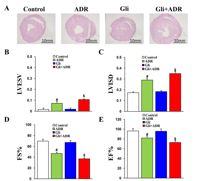
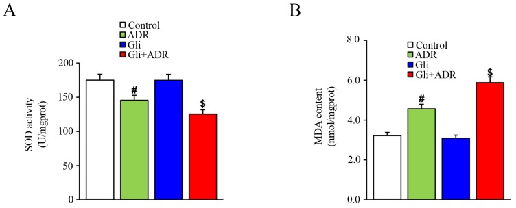
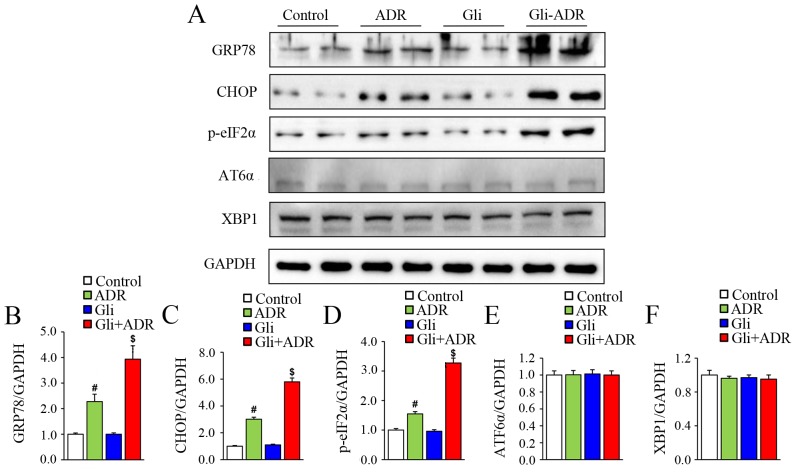
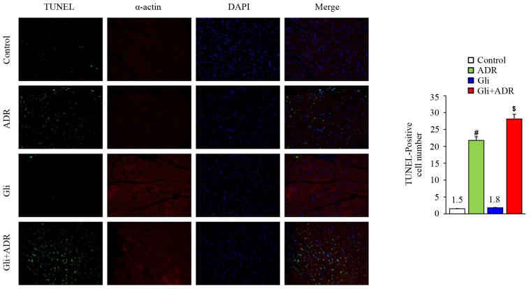
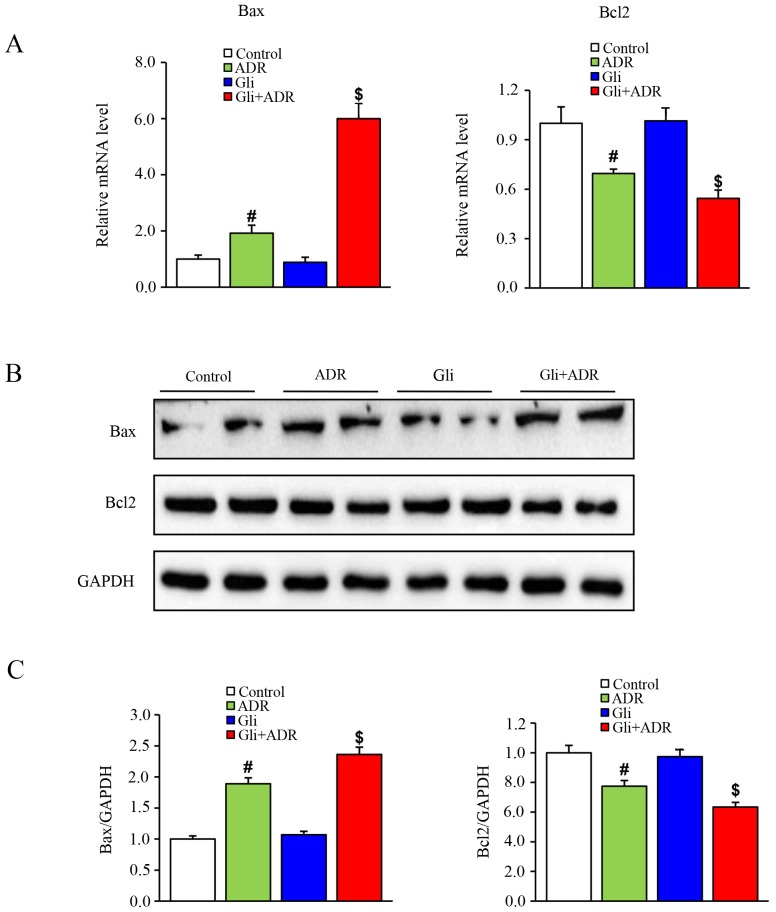
Adriamycin (ADR) is a chemotherapeutic drug used to treat tumors in a clinical setting. However, its use is limited by a side effect of cardiotoxicity. Glibenclamide (Gli), an inhibitor of mitochondrial ATP-dependent potassium (K-ATP) channels, blocks the cardioprotective effects of mitochondrial K-ATP channel openers and induces apoptosis in rodent pancreatic islet β-cell lines. However, little is known about the role of Gli in ADR-induced cardiotoxicity. The present study was designed to investigate the impact of Gli on ADR-induced cardiotoxicity in rats. A total of 60 male Sprague-Dawley rats were divided into the following 4 groups: i) Control; ii) Gli; iii) ADR; and iv) Gli+ADR (n=15 in each). The rats in the ADR and Gli+ADR groups were treated with ADR (intraperitoneal, 2.5 mg/kg/week) for 6 weeks. The rats in the Gli and Gli+ADR groups received Gli at a dose of 12 mg/kg/day via gastric lavage for 30 days from the eighth week of the study. Following the completion of Gli treatment, cardiac function was assessed by echocardiography, and the rats were sacrificed. The hearts were subsequently harvested for analysis. The rats in the ADR group demonstrated significantly impaired cardiac function and increased levels of oxidative stress, endoplasmic reticulum stress (ERS) and apoptosis in the heart compared with rats in the control and Gli groups (without ADR treatment). These abnormalities were exacerbated by Gli in the Gli+ADR group. Gli treatment decreased cardiac function and significantly increased oxidative stress, ERS and apoptosis levels in myocardial tissues in rats treated with ADR. The findings indicated that Gli triggers oxidative stress-induced ERS, and thus exacerbates ADR-induced cardiotoxicity in rats.

Keywords: adriamycin; apoptosis; endoplasmic reticulum stress; glibenclamide; oxidative stress.


Figures

Figure 1.
Figure 1.
Gli exacerbates ADR-induced impairments in cardiac function of rats. (A) Histological analysis of heart sections from the four groups (scale bar, 20 mm). The (B) LVESV, (C) LVISD, (D) FS% and (E) EF% data for the four groups. #P<0.05 vs. Control and Gli groups, $P<0.05 vs. ADR group. ADR, adriamycin; Gli, glibenclamide; LVESV, left ventricular end systolic volume; LVISD, left ventricular internal dimension systole; FS, fractional shortening; and EF, ejection fraction.
Figure 2.
Figure 2.
Effects of ADR and Gli on the expression levels of oxidative stress-related molecules. (A) SOD activity and (B) MDA content in myocardial tissue. #P<0.05 vs. Control and Gli groups, $P<0.05 vs. ADR group. ADR, adriamycin; Gli, glibenclamide; SOD, superoxide dismutase; and MDA, malondialdehyde.
Figure 3.
Figure 3.
Effects of Gli on ERS-related biomarker expression in ADR-treated rats. (A) Western blotting results for the protein expression levels of GRP78, CHOP, p-eIF2α, ATF6α and XBP1 in myocardial tissue and the quantified expression levels of (B) GRP78, (C) CHOP, (D), p-eIF2α, (E) ATF6α and (F) XBP1. #P<0.05 vs. Control and Gli groups, $P<0.05 vs. ADR group. ADR, adriamycin; Gli, glibenclamide; GRP78, glucose-regulated protein 78; p-eIF2α, phosphorylated eukaryotic translational initiation factor 2α; CHOP, C/EBP homologous protein; ATF6α, activating transcription actor 6α; and XBP1, X-box-binding protein-1.
Figure 4.
Figure 4.
TUNEL staining results. #P<0.05 vs. Control and Gli groups, $P<0.05 vs. ADR group. ADR, adriamycin; Gli, glibenclamide.
Figure 5.
Figure 5.
Effects of Gli on apoptosis-related biomarker expression in ADR-treated rats. (A) The quantified mRNA expression levels of Bax and Bcl-2 in myocardial tissue. (B) Western blotting results for the expression levels of Bax and Bcl-2 in myocardial tissue and (C) the quantified expression levels of Bax and Bcl-2. #P<0.05 vs. Control and Gli groups, $P<0.05 vs. ADR group. ADR, adriamycin; Gli, glibenclamide;

References

    1. Swain SM, Whaley FS, Ewer MS. Congestive heart failure in patients treated with doxorubicin: A retrospective analysis of three trials. Cancer. 2003;97:2869–2879. doi: 10.1002/cncr.11407. - DOI - PubMed
    1. Takemura G, Fujiwara H. Doxorubicin-induced cardiomyopathy From the cardiotoxic mechanisms to management. Prog Cardiovasc Dis. 2007;49:330–352. doi: 10.1016/j.pcad.2006.10.002. - DOI - PubMed
    1. Jones LW, Haykowsky MJ, Swartz JJ, Douglas PS, Mackey JR. Early breast cancer therapy and cardiovascular injury. J Am Coll Cardiol. 2007;50:1435–1441. doi: 10.1016/j.jacc.2007.06.037. - DOI - PubMed
    1. Octavia Y, Tocchetti CG, Gabrielson KL, Janssens S, Crijns HJ, Moens AL. Doxorubicin-induced cardiomyopathy: From molecular mechanisms to therapeutic strategies. J Mol Cell Cardiol. 2012;52:1213–1225. doi: 10.1016/j.yjmcc.2012.03.006. - DOI - PubMed
    1. Zhang S, Liu X, Bawa-Khalfe T, Lu LS, Lyu YL, Liu LF, Yeh ET. Identification of the molecular basis of doxorubicin-induced cardiotoxicity. Nat Med. 2012;18:1639–1642. doi: 10.1038/nm.2919. - DOI - PubMed