. 2018 Apr;15(4):3477-3482.
doi: 10.3892/etm.2018.5844. Epub 2018 Feb 7.

Slug inhibition increases radiosensitivity of nasopharyngeal carcinoma cell line C666-1

Affiliations

Slug inhibition increases radiosensitivity of nasopharyngeal carcinoma cell line C666-1

Hongxia Yang et al. Exp Ther Med. 2018 Apr.

Abstract

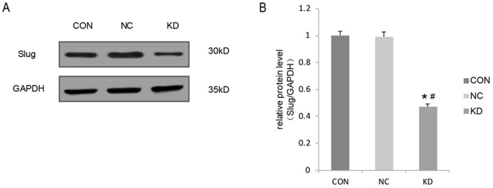
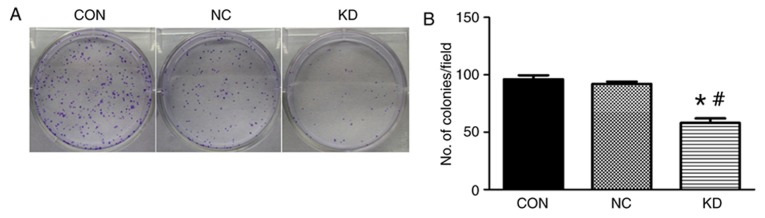
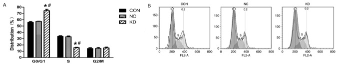
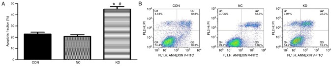
Slug is associated with the radioresistance of nasopharyngeal carcinoma (NPC) and the main current approach of treatment for NPC is radiotherapy. Hence, the aim of the current study was to determine the effect of Slug silencing on the radiosensitivity of NPC cells. Lentiviral-mediated transfection of Slug RNA interference (RNAi) in NPC cell line C666-1 was performed in vitro. Following Slug inhibition, its expression was detected using western blotting. A clonogenic survival assay and flow cytometry were then performed to evaluate the clonogenic cell survival, cell cycle distribution and apoptosis of C666-1 cells following irradiation. The results indicated that Slug RNAi decreased cell proliferation, and increased cell apoptosis and G0/G1 arrest. Thus, lentiviral-mediated transfection of Slug RNAi enhanced the radiosensitivity of the NPC cell line C666-1, and Slug may therefore be a potential target to improve radiotherapy in treatment of NPC and reduce the radioresistance of NPC.

Keywords: NPC; lentivirus; radiosensitivity; slug.


Figures

Figure 1.
Figure 1.
Lentiviral vectors encoding Slug RNAi or control RNAi were constructed and transfected into C666-1 cells, with untreated C666-1 cells serving as a control. (A) The expression of Slug was detected using western blotting. (B) Quantification of western blot results relative to GAPDH. Values are expressed as the mean ± standard deviation. All experiments were performed in triplicate. *P<0.05 vs. NC group; #P<0.05 vs. CON group. RNAi, RNA interference; CON, control group consisting of untreated C666-1 cells; NC, negative control consisting of C666-1 cells infected with pGCSIL-neg-shRNA-LV; KD, Slug RNAi group consisting of C666-1 cells infected with pGCSIL-Slug-shRNA-LV.
Figure 2.
Figure 2.
Clonogenic assay was used to observe the survival of C666-1 cells. (A) A clonogenic assay was used to detect the colony formation ability of cells following irradiation at 4 Gy. (B) Quantification of the number of colonies of C666-1 cells. Values are expressed as the mean ± standard deviation. All experiments were performed in triplicate. *P<0.05 vs. NC group; #P<0.05 vs. CON group. CON, control group consisting of untreated C666-1 cells; NC, negative control consisting of C666-1 cells infected with pGCSIL-neg-shRNA-LV; KD, Slug RNAi group consisting of C666-1 cells infected with pGCSIL-Slug-shRNA-LV.
Figure 3.
Figure 3.
Changes in the cell cycle following irradiation at 4 Gy in C666-1 cells treated with Slug RNAi. (A) Proportion of cells in the G0/G1 and S phases of the cell cycle in the NC, CON and KD groups. (B) C666-1 cell cycles were detected using flow cytometry analysis in the different groups. Values are expressed as the mean ± standard deviation. All experiments were performed in triplicate. *P<0.05 vs. NC group; #P<0.05 vs. CON group. RNAi, RNA interference; CON, control group consisting of untreated C666-1 cells; NC, negative control consisting of C666-1 cells infected with pGCSIL-neg-shRNA-LV; KD, Slug RNAi group consisting of C666-1 cells infected with pGCSIL-Slug-shRNA-LV; G0, resting phase; G1, gap 1 phase; S, synthesis phase; G2, gap 2 phase; M, mitotic phase.
Figure 4.
Figure 4.
Apoptosis ratio of C666-1 cells treated with Slug RNAi following irradiation at 4 Gy. (A) Percentage of apoptotic C666-1 cells in the CON, NC and KD groups. (B) Apoptosis was determined using flow cytometry analysis in the different groups. Values are expressed as the mean ± standard deviation. All experiments were performed in triplicate. *P<0.05 vs. NC group; #P<0.05 vs. NC group. RNAi, RNA interference; CON, control group consisting of untreated C666-1 cells; NC, negative control consisting of C666-1 cells infected with pGCSIL-neg-shRNA-LV; KD, Slug RNAi group consisting of C666-1 cells infected with pGCSIL-Slug-shRNA-LV; FITC, fluorescein isothiocyanate; PI, propidium iodide.

Similar articles

Cited by

References

    1. Cancer incidence in five continents. Volume VIII. IARC Sci Publ. 2002:1–781. - PubMed
    1. Chua MLK, Wee JTS, Hui EP, Chan ATC. Nasopharyngeal carcinoma. Lancet. 2016;387:1012–1024. doi: 10.1016/S0140-6736(15)00055-0. - DOI - PubMed
    1. Cui Q, Feng FT, Xu M, Liu WS, Yao YY, Xie SH, Li XZ, Ye ZL, Feng QS, Chen LZ, et al. Nasopharyngeal carcinoma risk prediction via salivary detection of host and Epstein-Barr virus genetic variants. Oncotarget. 2016;8:95066–95074. - PMC - PubMed
    1. Chua ML, Chan AT. Gemcitabine: A game changer in nasopharyngeal carcinoma. Lancet. 2016;388:1853–1854. doi: 10.1016/S0140-6736(16)31394-0. - DOI - PubMed
    1. Xiao WW, Han F, Lu TX, Chen CY, Huang Y, Zhao C. Treatment outcomes after radiotherapy alone for patients with early-stage nasopharyngeal carcinoma. Int J Radiat Oncol Biol Phys. 2009;74:1070–1076. doi: 10.1016/j.ijrobp.2008.09.008. - DOI - PubMed