

Semaphorin 3A promotes osteogenic differentiation in human alveolar bone marrow mesenchymal stem cells
- PMID: 29545873
- PMCID: PMC5841084
- DOI: 10.3892/etm.2018.5813
Semaphorin 3A promotes osteogenic differentiation in human alveolar bone marrow mesenchymal stem cells
Abstract
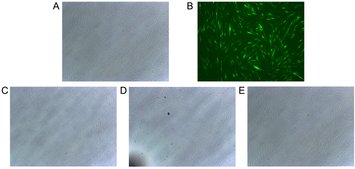
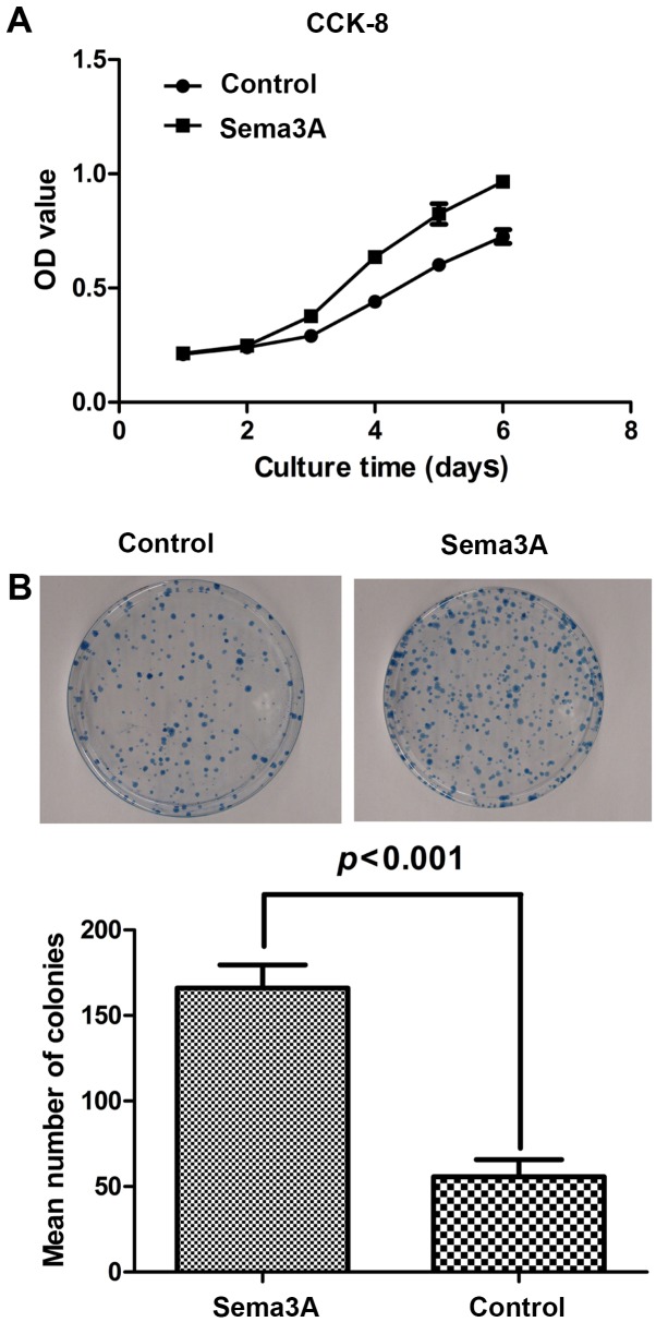
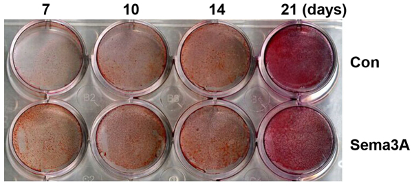
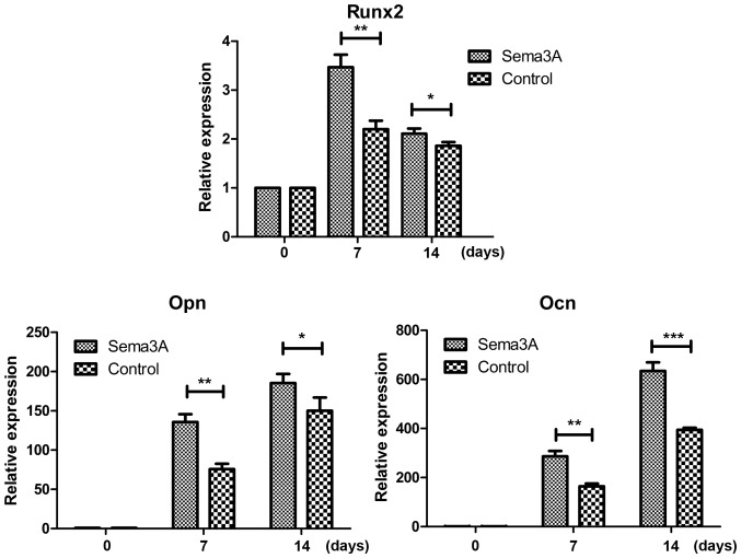
The aim of the present study was to investigate the role of Semaphorin 3A (Sema3A) in the osteogenic differentiation of human alveolar bone marrow mesenchymal stem cells (hABMMSCs). To investigate whether Sema3A affects hABMMSC proliferation and osteogenic differentiation, a stable Sema3A-overexpression cell line was generated by infection with the pAdCMV-SEMA3A-MCS-EGFP vector. Cell counting kit-8 and clone formation assays were performed to determine the proliferation ability of hABMMSCs, while cell osteogenic differentiation was assayed using Alizarin Red S staining. In addition, reverse transcription-quantitative polymerase chain reaction was employed to detect the mRNA expression level of osteogenesis-associated genes, Runt-related transcription factor 2 (Runx2), osteopontin (Opn) and osteocalcin (Ocn), during the osteogenic differentiation. The results revealed that, compared with the normal control group, the cell morphology of the infected cells was stable and no significant alterations were observed. Overexpression of Sema3A in hABMMSCs significantly increased the cell proliferation ability compared with the control group. Furthermore, the Alizarin Red S staining assay results indicated that the ossification process of hABMMSCs overexpressing Sema3A was evidently faster in comparison with that of the control group cells. Overexpression of Sema3A by pAdCMV-SEMA3A-MCS-EGFP infection also significantly increased the mRNA expression levels of the osteogenic marker genes Runx2, Opn and Ocn. In conclusion, Sema3A was observed to be a key positive regulator in hABMMSC osteogenic differentiation.
Keywords: human alveolar bone marrow mesenchymal stem cells; osteogenic differentiation; semaphorin 3A.
Figures
Similar articles
-
Semaphorin 3A promotes the osteogenic differentiation of rat bone marrow-derived mesenchymal stem cells in inflammatory environments by suppressing the Wnt/β-catenin signaling pathway.J Mol Histol. 2021 Dec;52(6):1245-1255. doi: 10.1007/s10735-020-09941-1. Epub 2021 Feb 10. J Mol Histol. 2021. PMID: 33566267
-
[Semaphorin 3A-stimulated bone marrow mesenchymal stem cells sheets promotes osteogenesis of type 2 diabetic rat].Zhonghua Kou Qiang Yi Xue Za Zhi. 2018 May 9;53(5):333-338. doi: 10.3760/cma.j.issn.1002-0098.2018.05.009. Zhonghua Kou Qiang Yi Xue Za Zhi. 2018. PMID: 29972992 Chinese.
-
The effects of Sema3A overexpression on the proliferation and differentiation of rat gingival mesenchymal stem cells in the LPS-induced inflammatory environment.Int J Clin Exp Pathol. 2019 Oct 1;12(10):3710-3718. eCollection 2019. Int J Clin Exp Pathol. 2019. PMID: 31933759 Free PMC article.
-
Semaphorin 3A-hypoxia inducible factor 1 subunit alpha co-overexpression enhances the osteogenic differentiation of induced pluripotent stem cells-derived mesenchymal stem cells in vitro.Chin Med J (Engl). 2020 Feb 5;133(3):301-309. doi: 10.1097/CM9.0000000000000612. Chin Med J (Engl). 2020. PMID: 31929360 Free PMC article.
-
Carbon monoxide releasing molecule‑3 promotes the osteogenic differentiation of rat bone marrow mesenchymal stem cells by releasing carbon monoxide.Int J Mol Med. 2018 Apr;41(4):2297-2305. doi: 10.3892/ijmm.2018.3437. Epub 2018 Jan 29. Int J Mol Med. 2018. PMID: 29393384
Cited by
-
Semaphorin 3A promotes the osteogenic differentiation of rat bone marrow-derived mesenchymal stem cells in inflammatory environments by suppressing the Wnt/β-catenin signaling pathway.J Mol Histol. 2021 Dec;52(6):1245-1255. doi: 10.1007/s10735-020-09941-1. Epub 2021 Feb 10. J Mol Histol. 2021. PMID: 33566267
-
Peripheral nerve-derived Sema3A promotes osteogenic differentiation of mesenchymal stem cells through the Wnt/β-catenin/Nrp1 positive feedback loop.J Cell Mol Med. 2024 Apr;28(8):e18201. doi: 10.1111/jcmm.18201. J Cell Mol Med. 2024. PMID: 38568078 Free PMC article.
-
Extracellular vesicles from deciduous pulp stem cells recover bone loss by regulating telomerase activity in an osteoporosis mouse model.Stem Cell Res Ther. 2020 Jul 17;11(1):296. doi: 10.1186/s13287-020-01818-0. Stem Cell Res Ther. 2020. PMID: 32680564 Free PMC article.
-
Neural regulation of mesenchymal stem cells in craniofacial bone: development, homeostasis and repair.Front Physiol. 2024 Jul 29;15:1423539. doi: 10.3389/fphys.2024.1423539. eCollection 2024. Front Physiol. 2024. PMID: 39135707 Free PMC article. Review.
-
The Role of Semaphorins and Their Receptors in Innate Immune Responses and Clinical Diseases of Acute Inflammation.Front Immunol. 2021 May 3;12:672441. doi: 10.3389/fimmu.2021.672441. eCollection 2021. Front Immunol. 2021. PMID: 34012455 Free PMC article. Review.
References
LinkOut - more resources
Full Text Sources
Other Literature Sources
Research Materials