

Berberine ameliorates neonatal necrotizing enterocolitis by activating the phosphoinositide 3-kinase/protein kinase B signaling pathway
- PMID: 29545879
- PMCID: PMC5841013
- DOI: 10.3892/etm.2018.5858
Berberine ameliorates neonatal necrotizing enterocolitis by activating the phosphoinositide 3-kinase/protein kinase B signaling pathway
Abstract
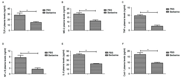
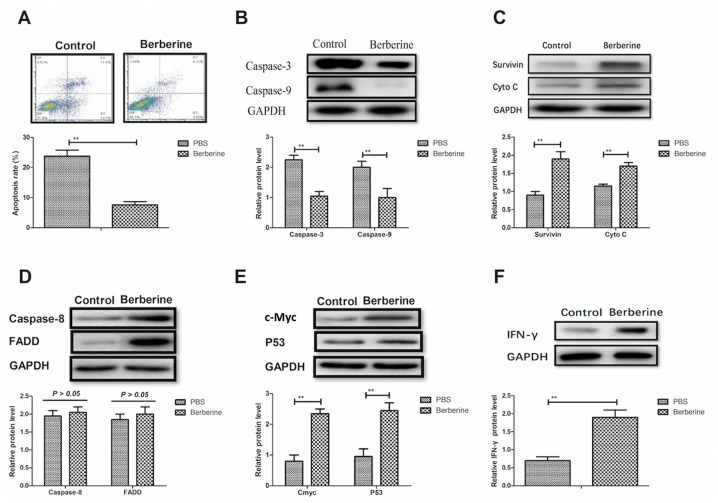
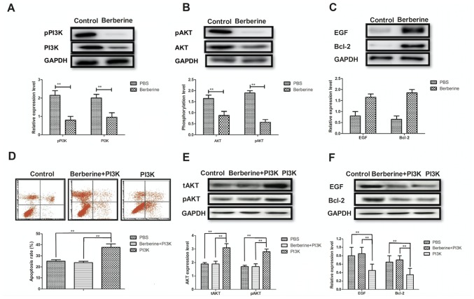
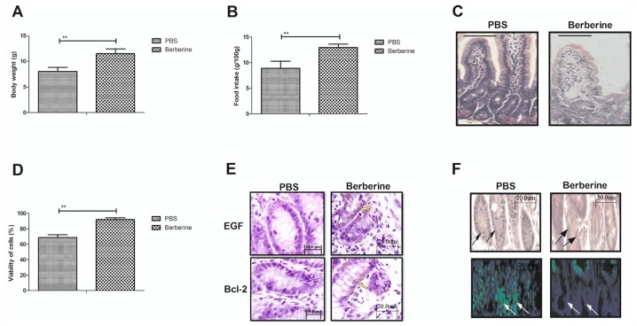
Neonatal necrotizing enterocolitis (NEC) is a severe acquired disease that predominantly affects the small intestine of neonates. NEC is caused by a combination of metabolic products, dysfunctions of the blood vessels, mucus and other unknown factors. Berberine may induce beneficial effects on necrotic and cardiovascular diseases due to its anti-inflammatory and anti-apoptotic effects on epithelial cells. In the present study, the therapeutic effects of berberine were investigated and the potential mechanisms by which it functions within a neonatal NEC mouse model were analyzed. Inflammation and levels of associated factors were measured in the serum of mice with NEC prior to and following treatment with berberine. Apoptotic rates in epithelial cells were analyzed following treatment with berberine. The expression of genes associated with apoptosis and apoptosis signaling were determined in epithelial cells in the small intestines of mice with NEC following treatment with berberine. The phosphoinositide 3-kinase (PI3K)/protein kinase B (AKT) signaling pathway was investigated in epithelial cells isolated from mice following treatment with either berberine or PBS. Histology and immunohistochemistry were used to determine the area of infarction and apoptosis. Body weight and food intake were measured to evaluate the physical effects of berberine on mice with NEC. The results indicated that berberine attenuated the inflammation caused by NEC in mice after 10 days of treatment. The apoptosis rate of epithelial cells isolated from experimental mice was decreased following berberine treatment. Western blot analysis indicated that the expression of the anti-apoptotic genes c-Myc and p53 were upregulated by berberine, whereas caspase-3 and -9 levels were downregulated in epithelial cells following treatment with berberine. In addition, the expression and phosphorylation levels of PI3K and AKT were downregulated in epithelial cells following treatment with berberine. An in vitro assay indicated that treatment with PI3K alone increased the expression of AKT and promoted the apoptosis of epithelial cells. Treatment with berberine markedly increased epidermal growth factor (EGF) and Bcl-2 expression levels, the activity of epithelial cells and decreased the infarction area of the small intestine. Accordingly, the body weight and food intake of mice with NEC were increased following berberine treatment. Therefore, the results of the present study demonstrate that berberine inhibits inflammation and apoptosis via the PI3K/AKT signaling pathway and may therefore attenuate the progression of NEC. These results suggest that berberine may be a potential therapeutic agent for the treatment of patients with NEC.
Keywords: apoptosis; berberine; inflammation; neonatal necrotizing enterocolitis; phosphoinositide 3 kinase/protein kinase B signaling pathway.
Figures
Similar articles
-
Vitamin A enhances PI3K/Akt signaling and mitigates enterocyte apoptosis in a mouse model of necrotizing enterocolitis.Pediatr Surg Int. 2025 Jan 8;41(1):61. doi: 10.1007/s00383-025-05963-6. Pediatr Surg Int. 2025. PMID: 39777515
-
Berberine inhibits the ischemia-reperfusion injury induced inflammatory response and apoptosis of myocardial cells through the phosphoinositide 3-kinase/RAC-α serine/threonine-protein kinase and nuclear factor-κB signaling pathways.Exp Ther Med. 2018 Feb;15(2):1225-1232. doi: 10.3892/etm.2017.5575. Epub 2017 Nov 27. Exp Ther Med. 2018. PMID: 29403554 Free PMC article.
-
Reversal of Mucin 1 Reduction-Induced Enterocyte Apoptosis by Retinoic Acid through the PI3K/AKT Signaling Pathway in an In vitro Model of Necrotizing Enterocolitis.Curr Mol Med. 2025 Jan 15. doi: 10.2174/0115665240337176241204070150. Online ahead of print. Curr Mol Med. 2025. PMID: 39819412
-
Anti-inflammatory mechanisms of bioactive milk proteins in the intestine of newborns.Int J Biochem Cell Biol. 2013 Aug;45(8):1730-47. doi: 10.1016/j.biocel.2013.04.028. Epub 2013 May 6. Int J Biochem Cell Biol. 2013. PMID: 23660296 Review.
-
Heparin-binding EGF-like growth factor (HB-EGF) and necrotizing enterocolitis.Semin Pediatr Surg. 2005 Aug;14(3):167-74. doi: 10.1053/j.sempedsurg.2005.05.005. Semin Pediatr Surg. 2005. PMID: 16084404 Review.
Cited by
-
Berberine: A Review of its Pharmacokinetics Properties and Therapeutic Potentials in Diverse Vascular Diseases.Front Pharmacol. 2021 Nov 3;12:762654. doi: 10.3389/fphar.2021.762654. eCollection 2021. Front Pharmacol. 2021. PMID: 35370628 Free PMC article. Review.
-
Targeting TREM1 Ameliorates Necrotizing Enterocolitis in Neonatal Mice by Inhibiting Endoplasmic Reticulum Stress and Regulating Macrophage Polarization.Dig Dis Sci. 2025 Jun 24. doi: 10.1007/s10620-025-09135-3. Online ahead of print. Dig Dis Sci. 2025. PMID: 40553398
-
Long-Term Phellodendri Cortex Supplementation in the Tiger Grouper (Epinephelus fuscoguttatus): Dual Effects on Intestinal Health Revealed by Transcriptome Analysis.Life (Basel). 2023 Dec 13;13(12):2336. doi: 10.3390/life13122336. Life (Basel). 2023. PMID: 38137937 Free PMC article.
-
Cytokine storm-calming property of the isoquinoline alkaloids in Coptis chinensis Franch.Front Pharmacol. 2022 Sep 6;13:973587. doi: 10.3389/fphar.2022.973587. eCollection 2022. Front Pharmacol. 2022. PMID: 36147356 Free PMC article. Review.
-
β-Carotene Attenuates Apoptosis and Autophagy via PI3K/AKT/mTOR Signaling Pathway in Necrotizing Enterocolitis Model Cells IEC-6.Evid Based Complement Alternat Med. 2022 Jun 17;2022:2502263. doi: 10.1155/2022/2502263. eCollection 2022. Evid Based Complement Alternat Med. 2022. PMID: 35754683 Free PMC article.
References
-
- Barak S, Riskin A, Kugelman A, Abend-Weinger M, Chistyakov I, Bader D. Necrotizing enterocolitis in a premature infant as the presenting symptom of familial dysautonomia in the neonatal period: Case report and review of the literature. Am J Perinatol. 2005;22:353–355. doi: 10.1055/s-2005-871525. - DOI - PubMed
LinkOut - more resources
Full Text Sources
Other Literature Sources
Research Materials
Miscellaneous