

Rapamycin promotes the anticancer action of dihydroartemisinin in breast cancer MDA-MB-231 cells by regulating expression of Atg7 and DAPK
- PMID: 29545903
- PMCID: PMC5840757
- DOI: 10.3892/ol.2018.8013
Rapamycin promotes the anticancer action of dihydroartemisinin in breast cancer MDA-MB-231 cells by regulating expression of Atg7 and DAPK
Abstract
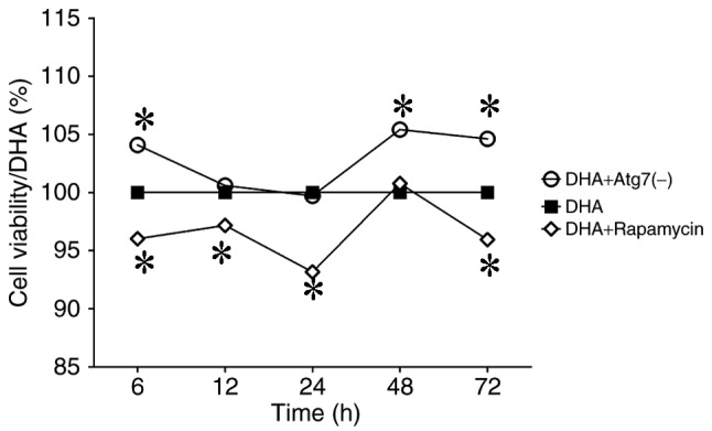
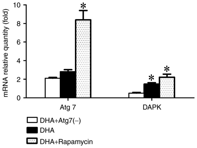
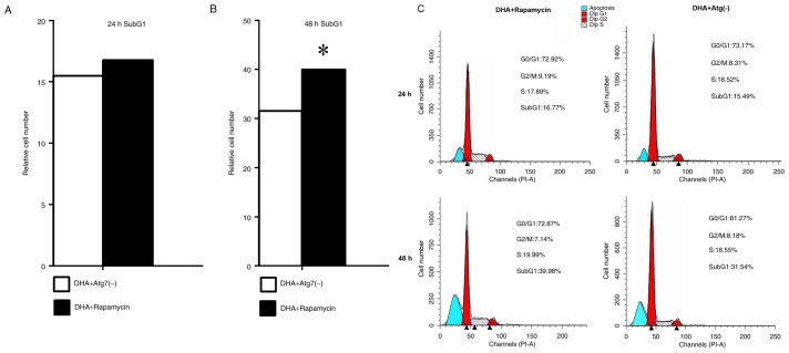
There is limited knowledge regarding the influence of autophagy on the anticancer effect of dihydroartemisinin (DHA). The present study aimed to investigate this influence within human breast cancer cells. Changes in cell viability, cell cycle distribution, apoptosis and associated genes were analyzed in MDA-MB-231 cells subjected to DHA following alteration in autophagy levels; the autophagy level was decreased following autophagy-related 7 (Atg7) knockdown or increased using rapamycin. The data indicated that rapamycin had the ability to notably enhance the anticancer effect of DHA on MDA-MB-231 cells. Autophagy induction may be key in mediating the anticancer effects of DHA, and rapamycin may regulate the death-associated protein kinase via the alteration of Atg7 expression, which would influence cell apoptosis. The present study presented a novel insight into enhancing the effectiveness of future treatment regimens for breast cancer using DHA.
Keywords: MDA-MB-231; autophagy; autophagy-related 7 small interfering RNA; breast cancer; dihydroartemisinin; rapamycin.
Figures
Similar articles
-
The pan-inhibitor of Aurora kinases danusertib induces apoptosis and autophagy and suppresses epithelial-to-mesenchymal transition in human breast cancer cells.Drug Des Devel Ther. 2015 Feb 17;9:1027-62. doi: 10.2147/DDDT.S74412. eCollection 2015. Drug Des Devel Ther. 2015. PMID: 25733818 Free PMC article.
-
The investigational Aurora kinase A inhibitor alisertib (MLN8237) induces cell cycle G2/M arrest, apoptosis, and autophagy via p38 MAPK and Akt/mTOR signaling pathways in human breast cancer cells.Drug Des Devel Ther. 2015 Mar 16;9:1627-52. doi: 10.2147/DDDT.S75378. eCollection 2015. Drug Des Devel Ther. 2015. PMID: 25834401 Free PMC article.
-
Effect of bis(hydroxymethyl) alkanoate curcuminoid derivative MTH-3 on cell cycle arrest, apoptotic and autophagic pathway in triple-negative breast adenocarcinoma MDA-MB-231 cells: An in vitro study.Int J Oncol. 2018 Jan;52(1):67-76. doi: 10.3892/ijo.2017.4204. Epub 2017 Nov 14. Int J Oncol. 2018. PMID: 29138806 Free PMC article.
-
Dihydroartemisinin and its anticancer activity against endometrial carcinoma and cervical cancer: involvement of apoptosis, autophagy and transferrin receptor.Singapore Med J. 2021 Feb;62(2):96-103. doi: 10.11622/smedj.2019138. Epub 2019 Nov 4. Singapore Med J. 2021. PMID: 31680182 Free PMC article.
-
Triptolide Decreases Cell Proliferation and Induces Cell Death in Triple Negative MDA-MB-231 Breast Cancer Cells.Biomolecules. 2018 Dec 5;8(4):163. doi: 10.3390/biom8040163. Biomolecules. 2018. PMID: 30563138 Free PMC article.
Cited by
-
Dihydroartemisinin: A Potential Drug for the Treatment of Malignancies and Inflammatory Diseases.Front Oncol. 2021 Oct 7;11:722331. doi: 10.3389/fonc.2021.722331. eCollection 2021. Front Oncol. 2021. PMID: 34692496 Free PMC article. Review.
-
Dihydroartemisinin Reduces Irradiation-Induced Mitophagy and Radioresistance in Lung Cancer A549 Cells via CIRBP Inhibition.Life (Basel). 2022 Jul 27;12(8):1129. doi: 10.3390/life12081129. Life (Basel). 2022. PMID: 36013308 Free PMC article.
-
Development of nanoscale drug delivery systems of dihydroartemisinin for cancer therapy: A review.Asian J Pharm Sci. 2022 Jul;17(4):475-490. doi: 10.1016/j.ajps.2022.04.005. Epub 2022 May 14. Asian J Pharm Sci. 2022. PMID: 36105316 Free PMC article. Review.
-
Dihydroartemisinin: A Potential Natural Anticancer Drug.Int J Biol Sci. 2021 Jan 16;17(2):603-622. doi: 10.7150/ijbs.50364. eCollection 2021. Int J Biol Sci. 2021. PMID: 33613116 Free PMC article. Review.
-
Anticancer Effect of Dihydroartemisinin via Dual Control of ROS-induced Apoptosis and Protective Autophagy in Prostate Cancer 22Rv1 Cells.Curr Pharm Biotechnol. 2024;25(10):1321-1332. doi: 10.2174/1389201024666230821155243. Curr Pharm Biotechnol. 2024. PMID: 37605406
References
-
- Plucinski MM, Dimbu PR, Macaia AP, Ferreira CM, Samutondo C, Quivinja J, Afonso M, Kiniffo R, Mbounga E, Kelley JS, et al. Efficacy of artemether-lumefantrine, artesunate-amodiaquine, and dihydroartemisinin-piperaquine for treatment of uncomplicated Plasmodium falciparum malaria in Angola, 2015. Malar J. 2017;16:62. doi: 10.1186/s12936-017-1712-4. - DOI - PMC - PubMed
-
- Thanh NV, Thuy-Nhien N, Tuyen NT, Tong NT, Nha-Ca NT, Dong LT, Quang HH, Farrar J, Thwaites G, White NJ, et al. Rapid decline in the susceptibility of Plasmodium falciparum to dihydroartemisinin-piperaquine in the south of Vietnam. Malar J. 2017;16:27. doi: 10.1186/s12936-017-1680-8. - DOI - PMC - PubMed
-
- Singh NP, Lai HC, Park JS, Gerhardt TE, Kim BJ, Wang S, Sasaki T. Effects of artemisinin dimers on rat breast cancer cells in vitro and in vivo. Anticancer Res. 2011;31:4111–4114. - PubMed
-
- Lai H, Nakase I, Lacoste E, Singh NP, Sasaki T. Artemisinin-transferrin conjugate retards growth of breast tumors in the rat. Anticancer Res. 2009;29:3807–3810. - PubMed
LinkOut - more resources
Full Text Sources
Other Literature Sources
Miscellaneous