Case Reports
. 2018 Aug;11(4):286-290.
doi: 10.1007/s12328-018-0848-2. Epub 2018 Mar 15.

A case of duodenal malignant lymphoma presenting as acute pancreatitis: systemic lupus erythematosus and immunosuppressive therapy as risk factors

Affiliations
Case Reports

A case of duodenal malignant lymphoma presenting as acute pancreatitis: systemic lupus erythematosus and immunosuppressive therapy as risk factors

Reiko Yamada et al. Clin J Gastroenterol. 2018 Aug.

Abstract

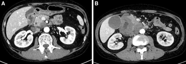
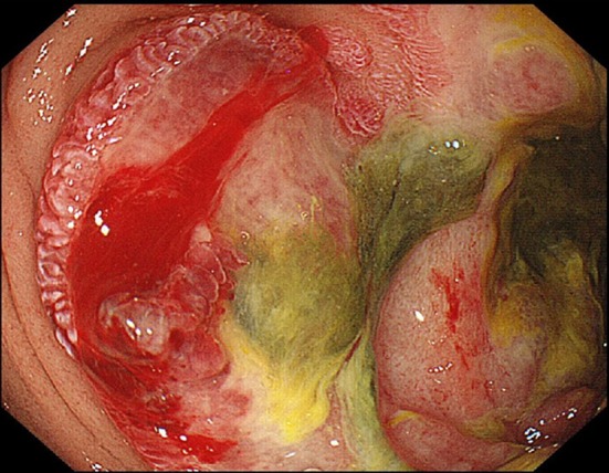
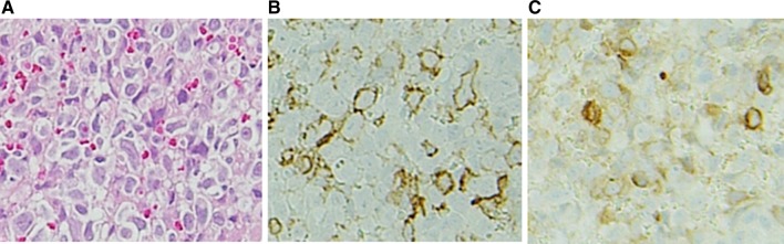
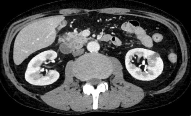
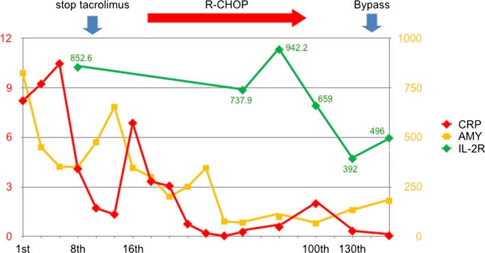
A 49-year-old man was admitted to our hospital with pancreatitis. He was diagnosed with systemic lupus erythematosus at 34 years of age and was being treated with oral tacrolimus (3 mg/day) and predonine (10 mg/day) for the past 15 months. The computed tomography (CT) scan showed the mass lesion had invaded the pancreatic head via thickening of the duodenal wall. Upper gastrointestinal endoscopy showed the all-round ulcerative lesion from the superior duodenal angle to the descending portion. Histological examination confirmed the diagnosis of diffuse large B cell lymphoma (DLBCL). Tacrolimus therapy was stopped due to the possibility of immunodeficiency-related lymphoproliferative disease; however, the lesion did not improve. Consequently, he was administered rituximab plus cyclophosphamide, doxorubicin, vincristine, and prednisone (R-CHOP). After six courses of R-CHOP therapy, a partial response was confirmed on CT. One month after the completion of chemotherapy, a gastrojejunal anastomosis was performed because of duodenal stenosis. He has since been well without recurrence. It was difficult to identify the risk factor for DLBCL; therefore, both the disease activity and immunosuppressive therapy should be taken into consideration as carrying a risk. In the present case, the symptom of pancreatitis enabled an early diagnosis of DLBCL.

Keywords: Duodenal malignant lymphoma; Immunosuppressive therapy-associated lymphoproliferative disorders; Pancreatitis; Systemic lupus erythematosus (SLE); Tacrolimus.


Conflict of interest statement

Conflict of interest

Reiko Yamada, Takashi Sakuno, Hiroyuki Inoue, Hiroshi Miura, Toshihumi Takeuchi, Yasunori Shiono, Hiroaki Okuse, Misaki Nakamura, Masaki Katsurahara, Yasuhiko Hamada, Kyosuke Tanaka, Noriyuki Horiki, and Yoshiyuki Takei declare that they have no conflict of interest.

Human rights

All procedures followed have been performed in accordance with the ethical standards laid down in the 1964 Declaration of Helsinki and its later amendments.

Informed consent

Informed consent was obtained from all patients for being included in the study.

Figures

Fig. 1
Fig. 1
The CT scan showed pancreatic head swelling and slight peripancreatic fluid effusion (a). The enhanced CT scan showed an all-round wall thickening of the descending portion of the duodenum and the ill-defined mass lesion invading the pancreatic head (b)
Fig. 2
Fig. 2
An upper gastrointestinal endoscopy showed the all-round ulcerative lesion over a large region from a superior duodenal angle (SDA) to the descending portion of duodenum. The well-demarcated lesion consisted of an ulcer with a regular elevated margin that had an auricle-like shape
Fig. 3
Fig. 3
Histological examination of the biopsy specimens confirmed the diagnosis of diffuse large B-cell lymphoma (DLBCL) (a); immunochemical staining was positive for CD20 (b) and CD79a (c)
Fig. 4
Fig. 4
After six courses of R-CHOP therapy, a 95.3% lesion rate of reduction was confirmed on CT, which was judged as a partial response
Fig. 5
Fig. 5
The chart shows that the initial laboratory tests indicated increased amylase (AMY; 823 IU/L) and C-reactive protein levels (CRP; 8.21 mg/dL). His abdominal pain was sustained on the day 8 after admission, although the inflammatory reaction and pancreatic enzyme levels were improving with fasting and infusion therapy: CRP = 4.09 mg/dL, and AMY = 350 IU/L. Tacrolimus was stopped on day 9, but the lesion continued to increase and his abdominal pain still persisted. Consequently, he was administered rituximab plus cyclophosphamide, doxorubicin, vincristine, and prednisone (R-CHOP) therapy from day 16. His symptoms improved and the laboratory data showed a good clinical course
Fig. 6
Fig. 6
An upper gastrointestinal endoscopy performed 1 month after the completion of chemotherapy revealed the scarring of duodenal ulcerative lesion and the high-grade duodenal stenosis

References

    1. Shnitser A, Halegoua-DeMarzio D, Loren DE. Primary pancreatic lymphoma presenting as acute pancreatitis. Gastroenterol Hepatol (N Y). 2016;12:456–458. - PMC - PubMed
    1. Nakamura S, Matsumoto T. Gastrointestinal lymphoma: recent advances in diagnosis and treatment. Digestion. 2013;87:182–188. doi: 10.1159/000350051. - DOI - PubMed
    1. Saif MW, Khubchandani S, Walczak M. Secondary pancreatic involvement by a diffuse large B-cell lymphoma presenting as acutepancreatitis. World J Gastroenterol. 2007;28:4909–4911. doi: 10.3748/wjg.v13.i36.4909. - DOI - PMC - PubMed
    1. Bernardeau M, Auroux J, Cavicchi M, et al. Secondary pancreatic involvement by diffuse large B-cell lymphoma presenting as acute pancreatitis: treatment and outcome. Pancreatology. 2002;2:427–430. doi: 10.1159/000065093. - DOI - PubMed
    1. Yadlapati S, Efthimiou P. Autoimmune/Inflammatory arthritis associated lymphomas: who is at risk? Biomed Res Int. 2016;2016:8631061. doi: 10.1155/2016/8631061. - DOI - PMC - PubMed

Publication types

MeSH terms