. 2018 Apr 10;57(14):2109-2120.
doi: 10.1021/acs.biochem.8b00065. Epub 2018 Mar 26.

CH···O Hydrogen Bonds Mediate Highly Specific Recognition of Methylated CpG Sites by the Zinc Finger Protein Kaiso

Affiliations

CH···O Hydrogen Bonds Mediate Highly Specific Recognition of Methylated CpG Sites by the Zinc Finger Protein Kaiso

Evgenia N Nikolova et al. Biochemistry. .

Abstract

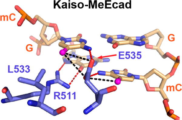
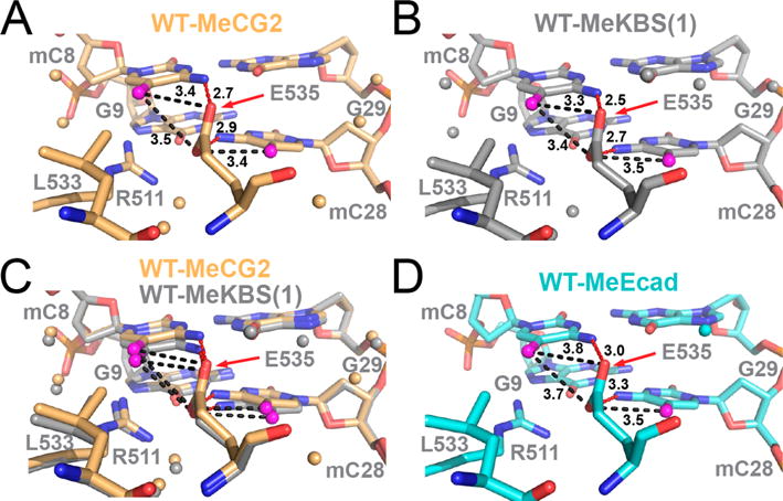
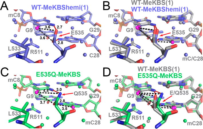
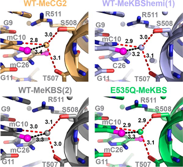
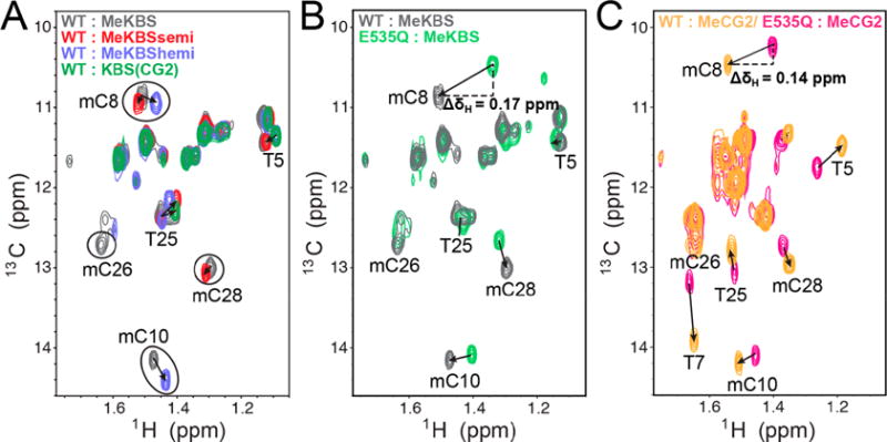
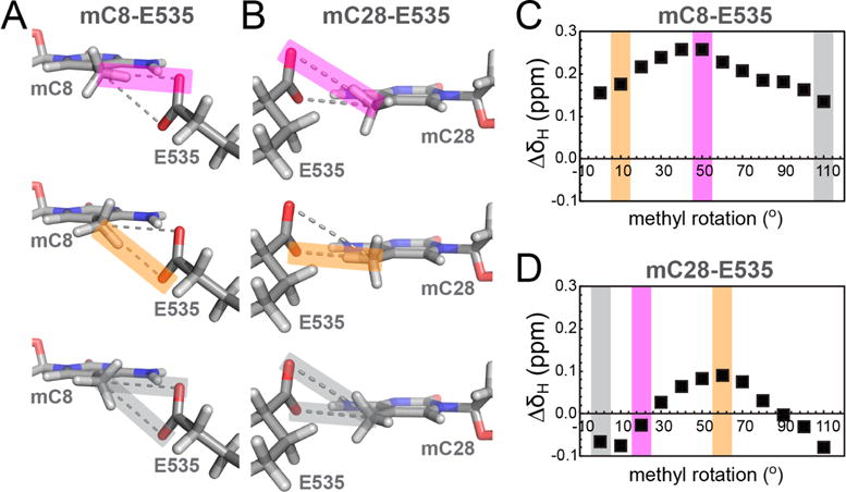
Many eukaryotic transcription factors recognize the epigenetic marker 5-methylcytosine (mC) at CpG sites in DNA. Despite their structural diversity, methyl-CpG-binding proteins (MBPs) share a common mode of recognition of mC methyl groups that involves hydrophobic pockets and weak hydrogen bonds of the CH···O type. The zinc finger protein Kaiso possesses a remarkably high specificity for methylated over unmethylated CpG sites. A key contribution to this specificity is provided by glutamate 535 (E535), which is optimally positioned to form multiple interactions with mCpG, including direct CH···O hydrogen bonds. To examine the role of E535 and CH···O hydrogen bonding in the preferential recognition of mCpG sites, we determined the structures of wild type Kaiso (WT) and E535 mutants and characterized their interactions with methylated DNA by nuclear magnetic resonance spectroscopy (NMR), X-ray crystallography, and in vitro protein-DNA binding assays. Our data show that Kaiso favors an mCpG over a CpG site by 2 orders of magnitude in affinity and that an important component of this effect is the presence of hydrophobic and CH···O contacts involving E535. Moreover, we present the first direct evidence for formation of a CH···O hydrogen bond between an MBP and 5-methylcytosine by using experimental (NMR) and quantum mechanical chemical shift analysis of the mC methyl protons. Together, our findings uncover a critical function of methyl-specific interactions, including CH···O hydrogen bonds, that optimize the specificity and affinity of MBPs for methylated DNA and contribute to the precise control of gene expression.


Conflict of interest statement

Notes

The authors declare no competing financial interest.

Figures

Figure 1
Figure 1
Mode of mCpG recognition in the methylated DNA (MeEcad) complex of Kaiso. Specific major groove interactions are shown between protein residues (blue) and a DNA mCpG site (wheat) in the crystal structure of Kaiso (Protein Data Bank entry 4F6N) bound to methylated DNA. mC methyl groups are colored magenta. Proposed canonical (red) and CH⋯O (black) hydrogen bonds are shown as dashed lines.
Figure 2
Figure 2
Sequences of DNA constructs used in this study and in the published studies for KBS and MeEcad. Shown are the central 10 bp around the core Kaiso-binding site (boxed). Residues are numbered as indicated for MeEcad, and methylated cytosines are colored red.
Figure 3
Figure 3
NMR spectra of complexes of Kaiso with methylated DNA. (A) Overlay of 1H–15N HSQC spectra of WT Kaiso bound to MeKBS (gray), MeKBShemi (blue), and MeKBSsemi (red). The inset shows a close-up of the E535 cross peak in these complexes, together with Kaiso–MeEcad (cyan) and Kaiso–MeCG2 (yellow) complexes. (B) Weighted average 1H–15N chemical shift differences [CSP = (0.1ΔδN2 + ΔδH2)1/2] between the backbone amide resonances of Kaiso in complex with MeKBS and MeKBShemi (blue) and in complex with MeKBS and MeKBSsemi (red), plotted as a function of residue number. (C) Overlay of 1H–15N HSQC spectra of Kaiso–MeKBS complexes of WT Kaiso (gray), E535Q (green), and E535A (orange). (D) CSPs between the backbone amide resonances of the MeKBS complexes of WT Kaiso and E535Q (green) and E535A (orange) as a function of residue number. Note that the y-axis scales in panels B and D differ by a factor of 2.
Figure 4
Figure 4
Structural comparison of the 5′ mCpG E535-binding interface in complexes of WT Kaiso with methylated DNA. (A) Complex of Kaiso with MeCG2 (yellow). (B) Complex of Kaiso with MeKBS (gray); MeKBS(1) refers to structure (1) reported in Table S2. (C) Overlay of the complexes of WT Kaiso with MeKBS and MeCG2. (D) Complex of Kaiso with MeEcad (cyan, PDB entry 4F6N). In each panel, the methyl carbon (C5A) of mC8 and mC28 is colored magenta and water molecules are shown as small spheres. Dashed lines show NH⋯O hydrogen bonds (red) between mC8(N4) and E535(Oε2), and between mC28(N4) and E535(Oε1), and potential CH⋯O hydrogen bonds (black) between mC8(C5A) and E535(Oε2) and between mC28(C5A) and E535(Oε1) (alternate CH⋯O hydrogen bonds with the E535 acceptor swapped are also possible but not shown for the sake of clarity). Donor–acceptor bond lengths are labeled in angstroms.
Figure 5
Figure 5
Effect of hemimethylation and E535Q mutation on the 5′ mCpG E535-binding interface in complexes of Kaiso with methylated DNA. (A) Complex of WT Kaiso with MeKBShemi (blue) (structure (1) reported in Table S2). (B) Overlay of the complexes of WT Kaiso with MeKBS and MeKBShemi. (C) Complex of Kaiso E535Q with MeKBS (green). (D) Overlay of the complexes of Kaiso WT and E535Q with MeKBS. In each panel, the methyl carbon (C5A) of mC8 and mC28 is colored magenta, water molecules are shown as small spheres, and donor–acceptor bond lengths are labeled in angstroms (see Figure 4 for a detailed description).
Figure 6
Figure 6
Conservation of the structure of the 3′ mCpG site in the complexes of MeCG2 (yellow), MeKBS (gray), and MeKBShemi (blue) with WT Kaiso and MeKBS with E535Q Kaiso (green). Potential water-mediated NH⋯O (red) and CH⋯O (black) hydrogen bonds between Kaiso T507/S508 and mC10 are shown by dashed lines, and distances are labeled in angstroms.
Figure 7
Figure 7
Direct NMR evidence for a CH⋯O hydrogen bond between Kaiso E535 and mCpG methyl groups in DNA. (A) Methyl region of the natural-abundance 1H–13C HMQC spectrum of DNA [MeKBS, gray; MeKBSsemi, red; MeKBShemi, blue; unmethylated KBS(CG2), green] in complex with WT Kaiso. (B) Overlay of the methyl region of the natural-abundance 1H–13C HMQC spectrum of MeKBS with Kaiso WT (gray) and E535Q (green). (C) Overlay of the methyl region of the natural-abundance 1H–13C HMQC spectrum of MeCG2 with Kaiso WT (yellow) and E535Q (magenta).
Figure 8
Figure 8
DFT chemical shift calculations for DNA mC methyl protons in Kaiso–MeKBS complexes. (A and B) Fragments from the crystal structure of the WT Kaiso–MeKBS complex showing DNA methyl group orientations that represent a nearly linear hydrogen bond alignment between mC8(C5A) (A) or mC28(C5A) (B) and E535(Oε2) (top, magenta), E535(Oε1) (middle, orange), or where on average the minimal effect on the proton chemical shift is observed, respectively (bottom, gray). (C and D) Representative plots of the difference in predicted methyl proton chemical shifts (ΔδH, ■) for mC8 and mC28, respectively, relative to the “no side chain” control. The chemical shift differences were calculated using DFT and are plotted as a function of counterclockwise methyl group rotation. The colored bands correspond to the methyl rotamers depicted in panel A or B.

References

    1. Li E. Chromatin modification and epigenetic reprogramming in mammalian development. Nat Rev Genet. 2002;3:662–673. - PubMed
    1. Reik W. Stability and flexibility of epigenetic gene regulation in mammalian development. Nature. 2007;447:425–432. - PubMed
    1. Kim JK, Samaranayake M, Pradhan S. Epigenetic mechanisms in mammals. Cell Mol Life Sci. 2009;66:596–612. - PMC - PubMed
    1. Klose RJ, Bird AP. Genomic DNA methylation: the mark and its mediators. Trends Biochem Sci. 2006;31:89–97. - PubMed
    1. Lister R, Ecker JR. Finding the fifth base: genome-wide sequencing of cytosine methylation. Genome Res. 2009;19:959–966. - PMC - PubMed

Publication types

Substances

LinkOut - more resources