

Plasticity of myeloid-derived suppressor cells in cancer
- PMID: 29547768
- PMCID: PMC5943174
- DOI: 10.1016/j.coi.2018.03.009
Plasticity of myeloid-derived suppressor cells in cancer
Abstract
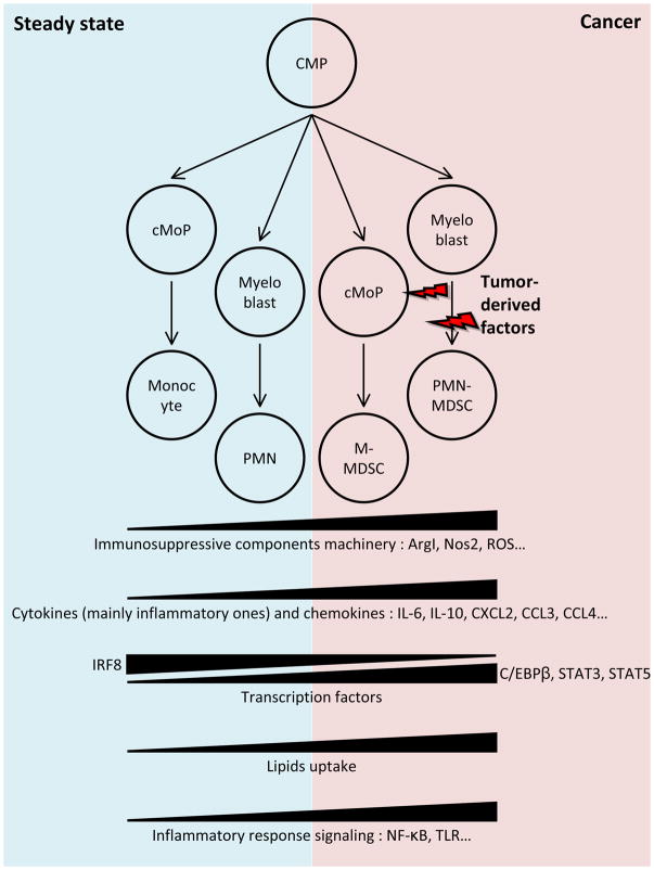
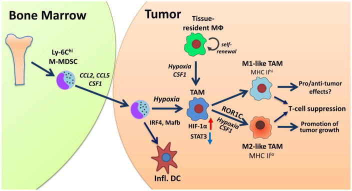
In recent years, myeloid-derived suppressor cells (MDSC) have emerged as one of the major inhibitors of immune effector cell function in cancer. MDSC represent a heterogeneous population of largely immature myeloid cells that are characterized by a pathological state of activation and display potent immune suppressive activity. Two major subsets of MDSC have been identified: monocytic (M-MDSC) and polymorphonuclear (PMN-MDSC). PMN-MSDC share phenotypic and morphologic features with neutrophils, whereas M-MDSC are similar to monocytes and are characterized by high plasticity. Differentiation of M-MDSC to macrophages and dendritic cells is shaped by tumor microenvironment. In recent years, the mechanisms of this process start to emerge.
Copyright © 2018 Elsevier Ltd. All rights reserved.
Figures
Similar articles
-
Myeloid-Derived Suppressor Cells: Immune-Suppressive Cells That Impair Antitumor Immunity and Are Sculpted by Their Environment.J Immunol. 2018 Jan 15;200(2):422-431. doi: 10.4049/jimmunol.1701019. J Immunol. 2018. PMID: 29311384 Free PMC article. Review.
-
Polymorphonuclear Myeloid-Derived Suppressor Cells Are Abundant in Peripheral Blood of Cancer Patients and Suppress Natural Killer Cell Anti-Tumor Activity.Front Immunol. 2022 Jan 18;12:803014. doi: 10.3389/fimmu.2021.803014. eCollection 2021. Front Immunol. 2022. PMID: 35116033 Free PMC article. Review.
-
Human neutrophils: Their role in cancer and relation to myeloid-derived suppressor cells.Semin Immunol. 2016 Apr;28(2):187-96. doi: 10.1016/j.smim.2016.03.018. Epub 2016 Apr 7. Semin Immunol. 2016. PMID: 27067179 Review.
-
Tumor promoting capacity of polymorphonuclear myeloid-derived suppressor cells and their neutralization.Int J Cancer. 2021 Nov 1;149(9):1628-1638. doi: 10.1002/ijc.33731. Epub 2021 Jul 17. Int J Cancer. 2021. PMID: 34224592 Review.
-
The Development and Homing of Myeloid-Derived Suppressor Cells: From a Two-Stage Model to a Multistep Narrative.Front Immunol. 2020 Oct 26;11:557586. doi: 10.3389/fimmu.2020.557586. eCollection 2020. Front Immunol. 2020. PMID: 33193327 Free PMC article. Review.
Cited by
-
Combination of transcriptome and Mendelian inheritance reveals novel prognostic biomarker of CTLA-4-related lncRNAs and protective role of nitrogen metabolism pathway in lung adenocarcinoma development.BMC Cancer. 2024 Aug 14;24(1):1009. doi: 10.1186/s12885-024-12777-7. BMC Cancer. 2024. PMID: 39143529 Free PMC article.
-
The role of myeloid-derived suppressor cells in gastrointestinal cancer.Cancer Commun (Lond). 2021 Jun;41(6):442-471. doi: 10.1002/cac2.12156. Epub 2021 Mar 27. Cancer Commun (Lond). 2021. PMID: 33773092 Free PMC article. Review.
-
Peri-operative monocyte count is a marker of poor prognosis in gastric cancer: increased monocytes are a characteristic of myeloid-derived suppressor cells.Cancer Immunol Immunother. 2019 Aug;68(8):1341-1350. doi: 10.1007/s00262-019-02366-0. Epub 2019 Jul 19. Cancer Immunol Immunother. 2019. PMID: 31324947 Free PMC article.
-
Myeloidcells in the immunosuppressive microenvironment in glioblastoma: The characteristics and therapeutic strategies.Front Immunol. 2023 Feb 27;14:994698. doi: 10.3389/fimmu.2023.994698. eCollection 2023. Front Immunol. 2023. PMID: 36923402 Free PMC article. Review.
-
Antitumor effects of targeting myeloid-derived suppressive cells.Transl Cancer Res. 2020 Sep;9(9):5787-5797. doi: 10.21037/tcr.2020.01.52. Transl Cancer Res. 2020. PMID: 35117939 Free PMC article. Review.
References
Publication types
MeSH terms
Substances
Grants and funding
LinkOut - more resources
Full Text Sources
Other Literature Sources
