

Acantholytic squamous cell carcinoma of the lung with marked lymphogenous metastases and high titers of myeloperoxidase-antineutrophil cytoplasmic antibodies: a case report
- PMID: 29548309
- PMCID: PMC5857100
- DOI: 10.1186/s12885-018-4218-8
Acantholytic squamous cell carcinoma of the lung with marked lymphogenous metastases and high titers of myeloperoxidase-antineutrophil cytoplasmic antibodies: a case report
Abstract
Background: Acantholytic squamous cell carcinoma (ASQCC), histologically characterized by intercellular bridge loosening, is recognized as a rare variant of squamous cell carcinoma (SQCC). ASQCC may demonstrate a worse prognosis than conventional SQCC. Pulmonary ASQCC is particularly rare; its biological behavior and prognostic data have not been reported.
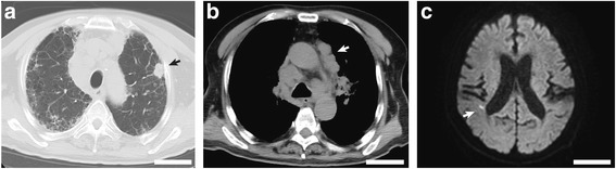
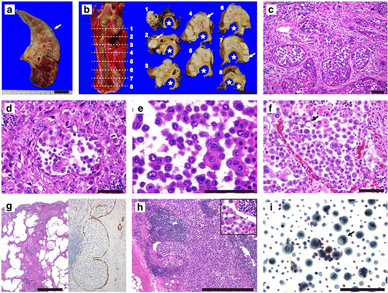
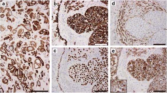
Case presentation: We report the clinical and autopsy findings of a 71-year-old Japanese man with pulmonary ASQCC. Pulmonary lesions, suggestive of idiopathic interstitial pneumonia, were radiologically observed 3 and 6 years prior to the patient's most recent hospitalization; however, the patient did not undergo further medical examinations. Upon being discovered unconscious, the patient was admitted to our hospital. Dehydration and lower limb muscle weakness were noted, as were laboratory findings of coagulation abnormalities and renal dysfunction. Computed tomography helped confirm a 21-mm peripheral nodule in the upper left lobe of the lung, with associated swollen lymph nodes in the bilateral hilar, mediastinal, and para-aortic regions. Brain and spinal lesions, suggestive of neurological disturbances, were not found. Small cell lung carcinoma was suspected, upon admission, but high serum levels of squamous cell carcinoma antigen and cytokeratin-19 fragments were present. Therefore, advanced lung cancer, possibly SQCC, was diagnosed. The patient was treated with best supportive therapy, and died one month after admission. Hypercalcemia and high serum levels of parathyroid hormone-related protein (PTHrP) and myeloperoxidase-antineutrophil cytoplasmic antibody (MPO-ANCA) titers were observed. Progressive renal insufficiency was absent due to improved renal function subsequent to hydration. An autopsy helped confirm the left lung tumor as an ASQCC associated with pulmonary lymphangitic carcinomatosis and multiple metastases in the lungs and lymph nodes. Skin lesions suggesting malignant tumors were absent. The metastatic lesions consisted largely of acantholytic tumor cells, and the lungs showed usual interstitial pneumonia pattern; vasculitis was absent.
Conclusions: This is the first reported case of pulmonary ASQCC resulting in an aggressive clinical course, with marked lymphogenous metastases and PTHrP-associated hypercalcemia. The high serum MPO-ANCA titers were clinicopathologically insignificant, but may have been related to the pulmonary interstitial lesion. Pulmonary ASQCC represents a highly malignant subset of lung cancer.
Keywords: Acantholytic squamous cell carcinoma; Hypercalcemia; Lymphogenous metastasis; Myeloperoxidase-antineutrophil cytoplasmic antibody; Parathyroid hormone–related protein; Small cell lung carcinoma; Squamous cell carcinoma.
Conflict of interest statement
Ethics approval and consent to participate
Our hospital’s ethics review board approved publication of this case report.
Consent for publication
Written informed consent for publication of this case was obtained from the patient’s son.
Competing interests
The authors declare that they have no competing interests.
Publisher’s Note
Springer Nature remains neutral with regard to jurisdictional claims in published maps and institutional affiliations.
Figures
Similar articles
-
Prognosis of pulmonary fibrosis presenting with a usual interstitial pneumonia pattern on computed tomography in patients with myeloperoxidase anti-neutrophil cytoplasmic antibody-related nephritis: a retrospective single-center study.BMC Pulm Med. 2019 Nov 1;19(1):194. doi: 10.1186/s12890-019-0969-5. BMC Pulm Med. 2019. PMID: 31675941 Free PMC article.
-
Radiologic and pathologic characteristics of myeloperoxidase-antineutrophil cytoplasmic antibody-associated interstitial lung disease: a retrospective analysis.Sarcoidosis Vasc Diffuse Lung Dis. 2019;36(3):195-201. doi: 10.36141/svdld.v36i3.8053. Epub 2019 May 1. Sarcoidosis Vasc Diffuse Lung Dis. 2019. PMID: 32476954 Free PMC article.
-
[A preliminary study of the significance of autoantibodies against light chain of myeloperoxidase on pulmonary damages in myeloperoxidase-antineutrophil cytoplasmic antibody associated vasculitis].Zhonghua Nei Ke Za Zhi. 2015 Jun;54(6):511-6. Zhonghua Nei Ke Za Zhi. 2015. PMID: 26359012 Chinese.
-
Acute tubulointerstitial nephritis associated with antineutrophil cytoplasmic antibody following cimetidine treatment: a case report.BMC Nephrol. 2021 Aug 30;22(1):294. doi: 10.1186/s12882-021-02502-y. BMC Nephrol. 2021. PMID: 34461843 Free PMC article. Review.
-
[Pulmonary mucosa-associated lymphoid tissue lymphoma concurrent with lung squamous cell carcinoma: a case report and literature review].Zhonghua Jie He He Hu Xi Za Zhi. 2020 Dec 12;43(12):1071-1076. doi: 10.3760/cma.j.cn112147-20200729-00859. Zhonghua Jie He He Hu Xi Za Zhi. 2020. PMID: 33333642 Review. Chinese.
Cited by
-
Clinical and Pathologic Presentation of Primary Ocular Surface Tumors among Zambians.Ocul Oncol Pathol. 2021 Mar;7(2):108-120. doi: 10.1159/000511610. Epub 2021 Jan 21. Ocul Oncol Pathol. 2021. PMID: 33869164 Free PMC article.
-
Acantholytic squamous cell carcinoma of the lung with pseudoglandular features: clinicopathologic study of 14 cases.Virchows Arch. 2025 Jul 3. doi: 10.1007/s00428-025-04149-8. Online ahead of print. Virchows Arch. 2025. PMID: 40608132
-
Atypical Histopathological Aspects of Common Types of Lung Cancer-Our Experience and Literature Review.Medicina (Kaunas). 2024 Jan 7;60(1):112. doi: 10.3390/medicina60010112. Medicina (Kaunas). 2024. PMID: 38256374 Free PMC article. Review.
-
Pulmonary Acantholytic Squamous Cell Carcinoma Mimicking Lepidic Pattern Adenocarcinoma.Turk Patoloji Derg. 2021;37(1):89-91. doi: 10.5146/tjpath.2019.01486. Turk Patoloji Derg. 2021. PMID: 32282054 Free PMC article. No abstract available.
-
Acantholytic Squamous Cell Carcinoma: a Diagnostic Pitfall on Cytology.Indian J Surg Oncol. 2023 Dec;14(4):963-967. doi: 10.1007/s13193-023-01811-y. Epub 2023 Sep 12. Indian J Surg Oncol. 2023. PMID: 38187856 Free PMC article.
References
-
- Weedon D, Morgan MB, Gross C, Nagore E, Yu LL. Squamous cell carcinoma. In: LeBoit PE, Burg G, Weedon D, Sarasin A, editors. World Health Organization classification of skin tumors. Lyon: International Agency for Research on Cancer; 2005. pp. 20–25.
-
- Reis-Filho JS, Lakhani SR, Gobbi H, Sneige N. Metaplastic carcinoma. In: Lakhani SR, Ellis LO, Schnitt SJ, Tan PH, van de Vijver MJ, editors. World Health Organization classification of the breast. Lyon: International Agency for Research on Cancer; 2012. pp. 48–52.
-
- Sloan P, Nylander K, Gale N, Reibel J, Hunter K, Salo T, et al. Malignant surface epithelial tumors. In: El-Naggar AK, Chan JKC, Grandis JR, Takata T, Slootweg PJ, et al., editors. World Health Organization classification of head and neck tumors. Lyon: International Agency for Research on Cancer; 2017. pp. 109–111.
-
- Cubilla AL, Dillner J, Amin MB, Moch H, Ayala A, Sanchez DF, et al. Malignant epithelial tumors. In: Moch H, Humphrey PA, Ulbright TM, Reuter VE, et al., editors. World Health Organization classification of tumors of the urinary system and male genital organs. Lyon: International Agency for Research on Cancer; 2016. pp. 262–276.
-
- Kirkham N, Aljefri K. Tumors and cysts of the epidermis. In: Elenitsas R, Rosenbach M, Murphy GF, Rubin AI, Xu X, editors. Lever’s histopathology of the skin. 11. Philadelphia: Lippincott Williams and Wilkins; 2015. pp. 969–1039.
Publication types
MeSH terms
Substances
LinkOut - more resources
Full Text Sources
Other Literature Sources
Medical
Research Materials
Miscellaneous
