. 2018 Apr:88:342-352.
doi: 10.1016/j.mcn.2018.03.005. Epub 2018 Mar 13.

CaBP1 regulates Cav1 L-type Ca2+ channels and their coupling to neurite growth and gene transcription in mouse spiral ganglion neurons

Affiliations

CaBP1 regulates Cav1 L-type Ca2+ channels and their coupling to neurite growth and gene transcription in mouse spiral ganglion neurons

Tian Yang et al. Mol Cell Neurosci. 2018 Apr.

Abstract

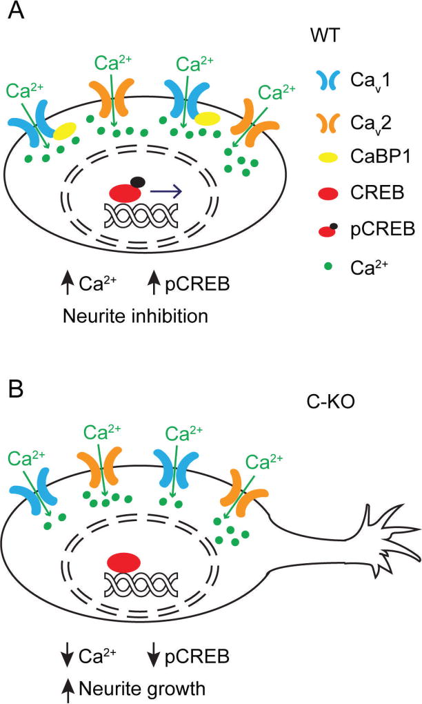
CaBP1 is a Ca2+ binding protein that is widely expressed in neurons in the brain, retina, and cochlea. In heterologous expression systems, CaBP1 interacts with and regulates voltage-gated Cav Ca2+ channels but whether this is the case in neurons is unknown. Here, we investigated the cellular functions of CaBP1 in cochlear spiral ganglion neurons (SGNs), which express high levels of CaBP1. Consistent with the role of CaBP1 as a suppressor of Ca2+-dependent inactivation (CDI) of Cav1 (L-type) channels, Cav1 currents underwent greater CDI in SGNs from mice lacking CaBP1 (C-KO) than in wild-type (WT) SGNs. The coupling of Cav1 channels to downstream signaling pathways was also disrupted in C-KO SGNs. Activity-dependent repression of neurite growth was significantly blunted and unresponsive to Cav1 antagonists in C-KO SGNs in contrast to WT SGNs. Moreover, Cav1-mediated Ca2+ signals and phosphorylation of cAMP-response element binding protein were reduced in C-KO SGNs compared to WT SGNs. Our findings establish a role for CaBP1 as an essential regulator of Cav1 channels in SGNs and their coupling to downstream pathways controlling activity-dependent transcription and neurite growth.

Keywords: Auditory; Ca(2+) channels; Neurite growth; Spiral ganglion.


Figures

Figure 1
Figure 1. Voltage-dependent activation is not affected but CDI is increased in C-KO SGNs
(A–B) I–V plots for ICa (A) and IBa (B) in WT and C-KO SGNs. Voltage protocol consisted of a 320-ms ramp from −45 mV to 70 mV. Black line and grey shading represent mean and SEM, respectively. (C) Representative traces for normalized currents evoked by a 1-s step to −10 mV (for IBa) or 0 mV (for ICa) for WT and C-KO SGNs. Scale bars, 0.5 s (horizontal); 200 pA for ICa (vertical). IBa was normalized to the scale of ICa. (D) For data obtained as in C, inactivation was measured as current amplitude at the end of the pulse normalized to the peak current amplitude (Ires/Ipk). (E) The difference in Ires/Ipk from D for ICa and IBa [ICa − IBa] was plotted for WT (n = 11 cells from 9 cultures from different litters) and C-KO SGNs (n = 9 cells from 8 cultures from different litters). Data were analyzed by unpaired t test. In D, E, points represent individual cells and bars represent mean ± SD; p values from unpaired t-tests are shown for each comparison. In D, t(18) = 3.207 for WT; t(15) = 5.894 for C-KO. In E, t(18) = 3.143.
Figure 2
Figure 2. Activity-dependent regulation of neuron survival is not altered in C-KO SGNs
Dissociated SGNs from WT or C-KO mice were maintained in culture for 4 h prior to depolarization with different [K+]o for 44 h prior to immunofluorescent labeling with NF200 antibodies. The control group was processed for NF200 labeling after the initial 4 h culture period. Survival (%) represents the number of SGNs at the end of depolarization period relative to the control for the indicated [K+]o. There was no significant difference in the effect of [K+]o on SGN survival in WT and C-KO cultures (F(1, 36) = 0.582, p = 0.450, by 2-way ANOVA, n = 4 cultures each).
Figure 3
Figure 3. Activity-dependent regulation of neurite growth is altered in C-KO SGNs
Dissociated SGNs from WT or C-KO mice were maintained in culture for 4 h prior to depolarization with different [K+]o for 44 h followed by immunofluorescent labeling with NF200 antibodies. (A,B) Representative images of WT (A) and C-KO (B) SGNs exposed to 30 mM or 80 mM [K+]o. Scale bar, 100 µm. Right, the percent of SGNs with neurites (relative to the total number of SGNs on the coverslip) at the end of the depolarization period is plotted for the indicated [K+]o. **, p < 0.01, ***, p < 0.001 compared to [K+]o = 5 mM by post-hoc Dunn’s multiple comparisons test. Each point represents result from different cultures (n = 5 cultures for WT, n = 7 cultures for C-KO; 3 mice were used per culture) and bars represent mean ± SD.
Figure 4
Figure 4. Neurite initiation but not rate of growth is enhanced in C-KO SGNs
SGNs were cultured for 12 h or 24 h in 30 mM [K+]o prior to NF200 labeling. (A, B) Representative images of WT and C-KO SGNs at the indicated time points. Scale bar, 100 µm. (C) The percent increase in SGNs with neurites at 24 h relative to 12 h is shown for WT and C-KO cultures (n = 5 cultures each, two replicates for each culture; t(8) = 3.102, p = 0.015, unpaired t test). Each point represents result from one culture and bars represent mean ± SD. (D, E) Distribution of neurite lengths (>10 µm) for SGN cultures after 12 h and 24 h for WT (D) and C-KO (E) cultures (n = 5 each, 106 and 216 WT SGNs at 12 h and 24 h, and 265 and 667 C-KO SGNs at 12 h and 24 h were analyzed). In D,E, results represent measurements on all SGNs in each culture.
Figure 5
Figure 5. Isradipine nullifies difference in neurite growth in WT and C-KO SGNs
SGNs were cultured in 30 mM [K+]o for 12 h in the presence of 0.1% DMSO or isradipine (0.1 µM or 10 µM) prior to NF200 labeling. (A,B) Representative images of WT and C-KO SGNs exposed to DMSO (control) or 0.1 µM isradipine (+ Isr). Scale bar, 100 µm. Right, the percentage of SGNs with neurites was measured as in Fig. 3 and shown for the indicated conditions. Isradipine had significant effects on WT SGNs (F(2, 8) = 14.63, p = 0.002 by ANOVA, n = 4 cultures/genotype), but not C-KO SGNs F(2, 9) = 0.63, p = 0.56 by ANOVA, n = 4 cultures/genotype). In A, p-value for the indicated comparison was determined by Bonferroni’s post-hoc test (t(8) = 5.374). Each point represents result from one culture and bars represent mean ± SD.
Figure 6
Figure 6. Bay K 8644 suppresses neurite growth and survival of WT but not C-KO SGNs
SGNs were cultured in 30 mM [K+]o for 44 h in the presence of 0.1% DMSO or Bay K 8644 (0.1 µM or 10 µM) prior to NF200 labeling. (A,B) Representative images of WT (A) and C-KO (B) SGN cultures exposed to DMSO (control) or 0.1 µM Bay K (+ Bay K). Scale bar, 100 µm. Right, the percentage of SGNs with neurites was measured as in Fig. 3, and the percent survival represents the number of SGNs at the end of the depolarization period normalized to the control. Bay K had a significant effect on neurite growth (F(2,9) = 7.57, p = 0.012 by ANOVA) and survival (F(2,9) = 20.5, p < 0.001, ANOVA), but not in C-KO cultures (F(2,9) = 3.91, p = 0.060, for neurite growth and F(2,9) = 0.72, p = 0.512 for neuron survival, ANOVA). p-values shown for the indicated comparisons were determined by Bonferroni’s test: #, t(9) = 3.52; ##, t(9) = 3.19; *, t(9) = 6.21; **, t(9) = 4.45. 4 WT and 4 C-KO cultures were analyzed. Each point represents result from one culture and bars represent mean ± SD.
Figure 7
Figure 7. Cav1-dependent phosphorylation of CREB is abolished in C-KO SGNs
(A,B) Representative images of WT and C-KO SGNs double-labeled with antibodies against pCREB (green) and NF200 (red) following a 15-min exposure to 5 or 30 mM [K+]o with or without isradipine (Isr, 10 µM). Scale bar, 10 µm. (C) Quantification of pCREB intensity in arbitrary units (a.u.). Results represent analyses of ~56 SGNs in 3 independent cultures. Each culture was prepared from 8 pups with 2 technical replicates per condition. 8–10 neurons were randomly selected from each replicate. Data are plotted as mean ± SEM. By Kruskal-Wallis and post-hoc Dunn’s test: #, compared to WT 5 mM, padj < 0.001 with mean rank difference of 171.9; ǂ, compared to C-KO 5 mM, padj < 0.001 with mean rank difference of 144.4; p-values are shown for the indicated comparisons. *, mean rank difference = 76.28; **, mean rank difference =104.9.
Figure 8
Figure 8. Cav1-mediated Ca2+ signals are reduced in C-KO SGNs
(A) Representative changes in GCaMP3 fluorescence (F/F0) in SGNs from WT or C-KO mice evoked by 80 mM [K+]o before and after application of isradipine. (B,C) Quantification of GCaMP3 fluorescent intensity changes in data obtained in A. p-values determined by Mann-Whitney test are indicated. U = 68.5 in B, U = 48.5 in C. Each point represents one region of interest and bars represent mean ± SD. F, maximum intensity; F0, intensity at time 0; Fb, baseline intensity. Data were collected from 4 WT and 4 C-KO cultures prepared from different litters. Each culture was prepared from 2–3 pups. For each experiment, 1–3 regions of interest were analyzed.
Figure 9
Figure 9. Cav1 signaling pathways that are altered in C-KO SGNs
(A) In WT SGNs, CaBP1 reduces CDI of Cav1 channels leading to increased Cav1-mediated signaling to pCREB and repression of neurite growth. (B) In C-KO SGNs, Cav1 channels undergo increased CDI and less coupling to pCREB and derepression of neurite growth.

Similar articles

Cited by

References

    1. Alseikhan BA, DeMaria CD, Colecraft HM, Yue DT. Engineered calmodulins reveal the unexpected eminence of Ca2+ channel inactivation in controlling heart excitation. Proc. Natl. Acad. Sci. U. S. A. 2002;99:17185–17190. - PMC - PubMed
    1. Audesirk G, Audesirk T, Ferguson C, Lomme M, Shugarts D, Rosack J, Caracciolo P, Gisi T, Nichols P. L-type calcium channels may regulate neurite initiation in cultured chick embryo brain neurons and N1E-115 neuroblastoma cells. Brain Res Dev Brain Res. 1990;55:109–120. - PubMed
    1. Bazzazi H, Ben Johny M, Adams PJ, Soong TW, Yue DT. Continuously tunable Ca2+ regulation of RNA-edited CaV1.3 channels. Cell rep. 2013;5:367–377. - PMC - PubMed
    1. Ben-Johny M, Yue DT. Calmodulin regulation (calmodulation) of voltage-gated calcium channels. J. Gen. Physiol. 2014;143:679–692. - PMC - PubMed
    1. Bengtson CP, Bading H. Nuclear calcium signaling. Adv Exp Med Biol. 2012;970:377–405. - PubMed

Publication types