

Template-based prediction of vigilance fluctuations in resting-state fMRI
- PMID: 29548849
- PMCID: PMC8328148
- DOI: 10.1016/j.neuroimage.2018.03.012
Template-based prediction of vigilance fluctuations in resting-state fMRI
Abstract
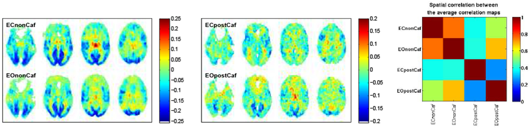
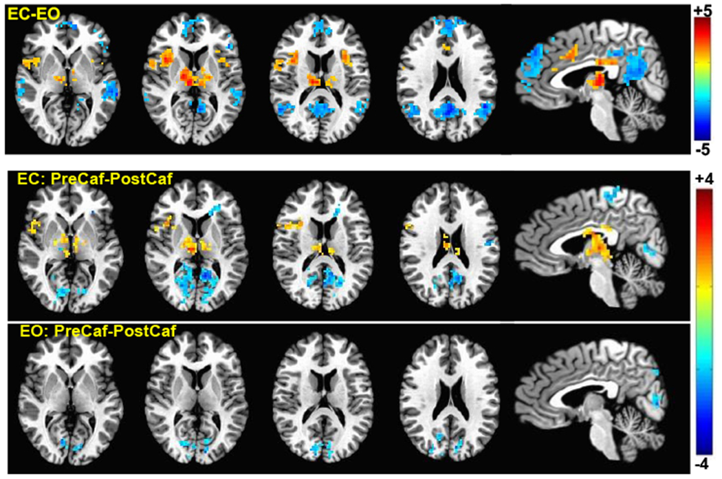
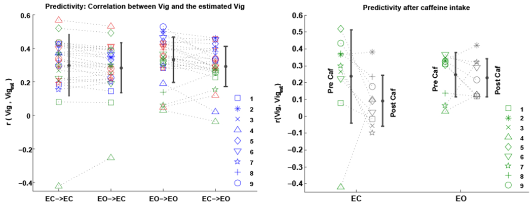
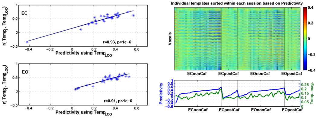
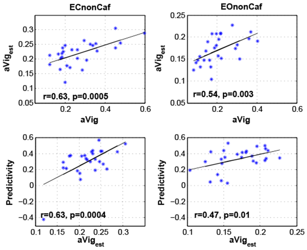
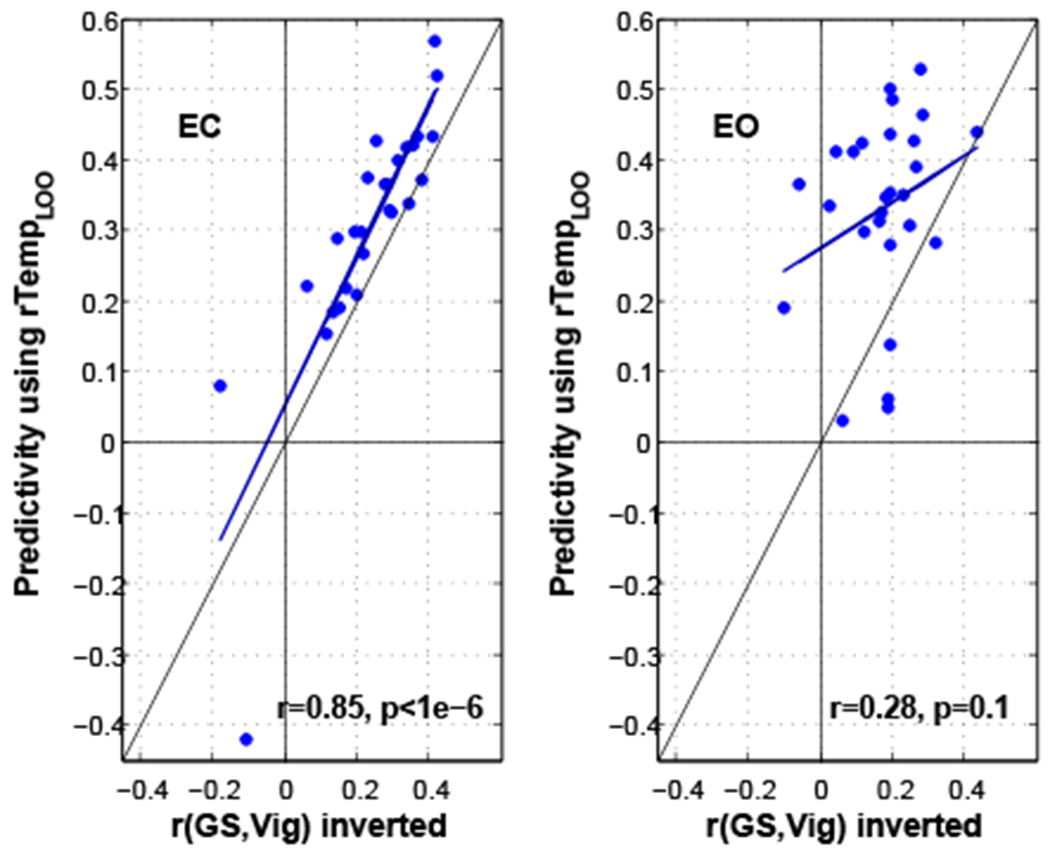
Changes in vigilance or alertness during a typical resting state fMRI scan are inevitable and have been found to affect measures of functional brain connectivity. Since it is not often feasible to monitor vigilance with EEG during fMRI scans, it would be of great value to have methods for estimating vigilance levels from fMRI data alone. A recent study, conducted in macaque monkeys, proposed a template-based approach for fMRI-based estimation of vigilance fluctuations. Here, we use simultaneously acquired EEG/fMRI data to investigate whether the same template-based approach can be employed to estimate vigilance fluctuations of awake humans across different resting-state conditions. We first demonstrate that the spatial pattern of correlations between EEG-defined vigilance and fMRI in our data is consistent with the previous literature. Notably, however, we observed a significant difference between the eyes-closed (EC) and eyes-open (EO) conditions, finding stronger negative correlations with vigilance in regions forming the default mode network and higher positive correlations in thalamus and insula in the EC condition when compared to the EO condition. Taking these correlation maps as "templates" for vigilance estimation, we found that the template-based approach produced fMRI-based vigilance estimates that were significantly correlated with EEG-based vigilance measures, indicating its generalizability from macaques to humans. We also demonstrate that the performance of this method was related to the overall amount of variability in a subject's vigilance state, and that the template-based approach outperformed the use of the global signal as a vigilance estimator. In addition, we show that the template-based approach can be used to estimate the variability across scans in the amplitude of the vigilance fluctuations. We discuss the benefits and tradeoffs of using the template-based approach in future fMRI studies.
Keywords: Caffeine; Eyes-closed; Eyes-open; Global signal; Resting state fMRI; Simultaneous EEG-fMRI; Vigilance.
Copyright © 2018 Elsevier Inc. All rights reserved.
Figures

Similar articles
-
Integration of Simultaneous Resting-State Electroencephalography, Functional Magnetic Resonance Imaging, and Eye-Tracker Methods to Determine and Verify Electroencephalography Vigilance Measure.Brain Connect. 2020 Dec;10(10):535-546. doi: 10.1089/brain.2019.0731. Epub 2020 Oct 28. Brain Connect. 2020. PMID: 33112650 Free PMC article.
-
The amplitude of the resting-state fMRI global signal is related to EEG vigilance measures.Neuroimage. 2013 Dec;83:983-90. doi: 10.1016/j.neuroimage.2013.07.057. Epub 2013 Jul 27. Neuroimage. 2013. PMID: 23899724 Free PMC article.
-
Differences in the resting-state fMRI global signal amplitude between the eyes open and eyes closed states are related to changes in EEG vigilance.Neuroimage. 2016 Jan 1;124(Pt A):24-31. doi: 10.1016/j.neuroimage.2015.08.053. Epub 2015 Aug 29. Neuroimage. 2016. PMID: 26327245
-
Endogenous brain oscillations and related networks detected by surface EEG-combined fMRI.Hum Brain Mapp. 2008 Jul;29(7):762-9. doi: 10.1002/hbm.20600. Hum Brain Mapp. 2008. PMID: 18465797 Free PMC article. Review.
-
Mapping cognitive and emotional networks in neurosurgical patients using resting-state functional magnetic resonance imaging.Neurosurg Focus. 2020 Feb 1;48(2):E9. doi: 10.3171/2019.11.FOCUS19773. Neurosurg Focus. 2020. PMID: 32006946 Free PMC article. Review.
Cited by
-
Inter-relationships between changes in stress, mindfulness, and dynamic functional connectivity in response to a social stressor.Sci Rep. 2022 Feb 14;12(1):2396. doi: 10.1038/s41598-022-06342-0. Sci Rep. 2022. PMID: 35165343 Free PMC article.
-
Functional MRI signals exhibit stronger covariation with peripheral autonomic measures as vigilance decreases.Imaging Neurosci (Camb). 2024 Sep 13;2:imag-2-00287. doi: 10.1162/imag_a_00287. eCollection 2024. Imaging Neurosci (Camb). 2024. PMID: 40800367 Free PMC article.
-
State-related neural influences on fMRI connectivity estimation.Neuroimage. 2021 Dec 1;244:118590. doi: 10.1016/j.neuroimage.2021.118590. Epub 2021 Sep 21. Neuroimage. 2021. PMID: 34560268 Free PMC article. Review.
-
Vigilance associates with the low-dimensional structure of fMRI data.Neuroimage. 2023 Feb 15;267:119818. doi: 10.1016/j.neuroimage.2022.119818. Epub 2022 Dec 16. Neuroimage. 2023. PMID: 36535323 Free PMC article.
-
Altered thalamic connectivity in insomnia disorder during wakefulness and sleep.Hum Brain Mapp. 2021 Jan;42(1):259-270. doi: 10.1002/hbm.25221. Epub 2020 Oct 13. Hum Brain Mapp. 2021. PMID: 33048406 Free PMC article.
References
-
- AASM, 2009. The AASM Manual for the Scoring of Sleep and Associated Events: Rules, Terminology and Technical Specification.
-
- Allen P, Josephs O, Turner R, August. 2000. A method for removing imaging artifact from continuous EEG recorded during functional MRI. Neuroimage 12, 230–239. - PubMed
-
- Altmann A, Schröter MS, Spoormaker VI, Kiem SA, Jordan D, Ilg R, Bullmore ET, Greicius MD, Czisch M, Sämann PG, January 2016. Validation of non-rem sleep stage decoding from resting state fmri using linear support vector machines. Neuroimage 125, 544–555. - PubMed
-
- Barry R, Clarke A, Johnstone S, Magee C, Rushby J, 2007. EEG differences between eyes-closed and eyes-open resting conditions. Clin Neurophysiol. 118, 2765–2773. - PubMed
-
- Braboszcz C, Delorme A, 2011. Lost in thoughts: neural markers of low alertness during mind wandering. Neuroimage 54, 3040–7. - PubMed
Publication types
MeSH terms
Grants and funding
LinkOut - more resources
Full Text Sources
Other Literature Sources
