. 2018 Mar 16;8(1):4664.
doi: 10.1038/s41598-018-22959-6.

Sites associated with Kalydeco binding on human Cystic Fibrosis Transmembrane Conductance Regulator revealed by Hydrogen/Deuterium Exchange

Affiliations

Sites associated with Kalydeco binding on human Cystic Fibrosis Transmembrane Conductance Regulator revealed by Hydrogen/Deuterium Exchange

Laura J Byrnes et al. Sci Rep. .

Abstract

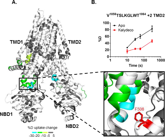
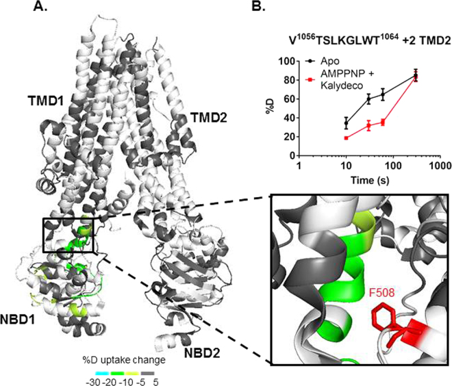
Cystic Fibrosis (CF) is caused by mutations in the Cystic Fibrosis Transmembrane Conductance Regulator (CFTR). Mutations associated with CF cause loss-of-function in CFTR leading to salt imbalance in epithelial tissues. Kalydeco (also called VX-770 or ivacaftor) was approved for CF treatment in 2012 but little is known regarding the compound's interactions with CFTR including the site of binding or mechanisms of action. In this study we use hydrogen/deuterium exchange (HDX) coupled with mass spectrometry to assess the conformational dynamics of a thermostabilized form of CFTR in apo and ligand-bound states. We observe HDX protection at a known binding site for AMPPNP and significant protection for several regions of CFTR in the presence of Kalydeco. The ligand-induced changes of CFTR in the presence of Kalydeco suggest a potential binding site.


Conflict of interest statement

The authors declare no competing interests.

Figures

Figure 1
Figure 1
FL hCFTRTS purification and activity. (A) Representative chromatograms for dephosphorylated (red) and PKA phosphorylated (blue) hCFTRTS run over a Superose 6 10/300 SEC column as a final purification step. (B) hCFTRTS after purification (no phosphatase or kinase treatment in lane (1), post λPP treatment in lane (2), and post PKA treatment in lane (3) was run on stain-free Bio-rad SDS-PAGE and imaged (left image). The same gel was then stained using Pro-Q Diamond Phosphoprotein Stain to confirm phosphorylation state after λPP and PKA treatment. The full-length gel images are shown in Figure S1B and C. (C) ATPase activity of purified dephosphorylated (red) and phosphorylated (blue) hCFTRTS at 0.5 µM in the presence of 5 mM ATP was measured using the PK/LDH coupled enzyme assay.
Figure 2
Figure 2
HDX profile of apo hCFTRTS. (A) HDX data from the heat map (see Figure S5) is indicated on the 3D cryo-EM structure of human CFTR (PDB 5UAK). The R domain is not shown in the cyro-EM structure since it was not fully resolved. As shown in the key, a color gradient is used to represent average deuterium uptake values across all 6 time points ranging from 10 s to 3600 s. White indicates regions that were not detected for every replicate at every time point in the HDX experiment. (BD) Representative deuterium uptake time-course curves are shown for peptides from RD (B), TMD2 (C) and NBD1 (D). Data represent the means ± SD (n = 3). Charge state of the detected peptide ion is also noted.
Figure 3
Figure 3
Stabilization of hCFTRTS upon AMPPNP binding. (A) HDX perturbation data mapped to the cryo-EM structure (PDB 5UAK). As shown in the key, a color gradient is used to represent the average deuterium uptake differences across all 6 time points between the apo and AMPPNP bound states of hCFTRTS. White indicates regions that were not detected for every replicate at every time point in the HDX experiments. NBD1 is rotated and zoomed in to show the stabilized peptides. Conserved motifs are highlighted as follows: Walker A sidechains (sticks), signature sequence (brown), and Walker B (red). Estimated location of ATP (magenta, sticks) and magnesium (orange, ball) are shown based on alignment with NBD1 crystal structure (2BBO). (B) Deuterium uptake time-course plots of a peptide from the Walker A motif in both apo and AMPPNP bound states. Data represent the means ± SD (n = 3). Charge state of the peptide is also noted.
Figure 4
Figure 4
hCFTRTS conformational changes upon Kalydeco binding. (A) HDX perturbation data mapped to the cryo-EM structure (PDB 5UAK). As shown in the key, a color gradient is used to represent the average deuterium uptake differences across all 6 time points between the apo and Kalydeco bound states of hCFTRTS. White indicates regions that were not detected for every replicate at every time point in the HDX experiments. The ICL4 region was zoomed in to show the peptides stabilized by Kalydeco binding. F508 was highlighted in red and represented as sticks. (B) Deuterium uptake time-course curves of the TMD2 peptide which showed protection upon ligand binding. Data represent the means ± SD (n = 2–3). Charge state of the peptide ion is also noted.
Figure 5
Figure 5
hCFTRTS conformational changes when both AMPPNP and Kalydeco were bound. (A) HDX perturbation data mapped to the cryo-EM structure (PDB 5UAK). As shown in the key, a color gradient is used to represent the average deuterium uptake differences across all 6 time points between the apo and ligand bound states of hCFTRTS. White indicates regions that were not detected for every replicate at every time point in the HDX experiments. The ICL4 region was zoomed in to show the protected peptides. F508 was highlighted in red and represented as sticks. (B) Deuterium uptake time-course curves of the TMD2 peptide which showed protection upon ligand binding. Data represent the means ± SD (n = 2–3). Charge state of the peptide ion is also noted.

Similar articles

Cited by

References

    1. Collins FS. Cystic fibrosis: molecular biology and therapeutic implications. Science. 1992;256:774–779. doi: 10.1126/science.1375392. - DOI - PubMed
    1. Davis PB. Cystic fibrosis since 1938. Am J Respir Crit Care Med. 2006;173:475–482. doi: 10.1164/rccm.200505-840OE. - DOI - PubMed
    1. Van Goor F, et al. Rescue of CF airway epithelial cell function in vitro by a CFTR potentiator, VX-770. Proc Natl Acad Sci USA. 2009;106:18825–18830. doi: 10.1073/pnas.0904709106. - DOI - PMC - PubMed
    1. Moran O. The gating of the CFTR channel. Cell Mol Life Sci. 2017;74:85–92. doi: 10.1007/s00018-016-2390-z. - DOI - PMC - PubMed
    1. Cheng SH, et al. Phosphorylation of the R domain by cAMP-dependent protein kinase regulates the CFTR chloride channel. Cell. 1991;66:1027–1036. doi: 10.1016/0092-8674(91)90446-6. - DOI - PubMed

MeSH terms

LinkOut - more resources